The Axolotl Mystery: Why This Salamander Can Regrow Anything
When an axolotl loses a limb, it doesn’t form a scar. Instead, the wound closes within hours, nearby cells reverse their specialization, and a dome of rapidly dividing progenitor cells called a blastema builds the entire limb from scratch — bone, cartilage, muscle, nerves, blood vessels, even fingerprints of texture — in about 60 days. The axolotl does this using many of the same genes humans carry. The difference isn’t in the hardware. It’s in the software.
How does the axolotl regrow entire limbs? Cut off an axolotl’s leg, and something happens that should be impossible. The wound doesn’t scar. The stump doesn’t seal over and forget. Instead, cells at the wound site do something that no adult human cell can do on its own — they go backward.
Mature muscle cells, cartilage cells, connective tissue cells — cells that were done growing, done dividing, done specializing — strip away their adult identity, re-enter the cell cycle, and begin building a brand-new limb from nothing. Bone. Tendons. Nerves. Blood vessels. Skin. Every tissue, in the right place, in the right proportion, with full function restored.
And here’s the part that keeps biologists awake at night: axolotl regeneration uses many of the same genes that exist in the human genome. We have the genetic toolkit. We just don’t use it.
In the next 18 minutes, I’ll walk you through exactly how axolotl limb regeneration works — from the first seconds after injury to the final rebuilt finger — and why this process matters for the future of human medicine.
Key takeaways
- Axolotls can regenerate entire limbs, portions of the brain, spinal cord, heart, lungs, jaw, retina, and ovary — repeatedly, throughout life.
- The secret is the blastema — a mass of progenitor cells that forms at the wound site instead of scar tissue.
- Mature cells dedifferentiate (reverse their specialization) but retain memory of their tissue type — muscle makes muscle, bone makes bone.
- The process relies on suppressing the scarring response — something humans do the opposite of.
- Axolotl macrophages send pro-regenerative signals instead of the pro-inflammatory signals that drive human scarring.
- Positional memory is encoded by retinoic acid gradients and genes like Hand2 — telling the blastema which body part to rebuild.
- Axolotls are Critically Endangered — perhaps only 50–1,000 remain in the wild in Lake Xochimilco, Mexico.
- The axolotl genome is 32 billion base pairs — 10× the human genome — the largest genome ever sequenced.
- What Can an Axolotl Actually Regenerate?
- Step 1 — Wound Healing Without Scarring
- Step 2 — Dedifferentiation: Cells Going Backward
- Step 3 — The Blastema: Biology’s Construction Site
- Step 4 — How Does It Know What to Build?
- Step 5 — Rebuilding Tissue by Tissue
- The Nerve Dependency — No Nerves, No Regeneration
- The Macrophage Switch — Inflammation vs. Regeneration
- The Largest Genome Ever Sequenced
- Neoteny — The Salamander That Never Grows Up
- Why Humans Scar Instead of Regenerate
- What Axolotl Regeneration Means for Human Medicine
- Critically Endangered — The Paradox of the Axolotl
- Axolotl Limb Regeneration — Day-by-Day Timeline
- Frequently Asked Questions
- References
1. What Can an Axolotl Actually Regenerate?
Before we get into how axolotl regeneration works, let’s establish the sheer scope of what these animals can do. The axolotl (Ambystoma mexicanum) is the most regenerative vertebrate known to science.
| Structure | Regeneration quality | Can humans do this? |
|---|---|---|
| All four limbs | Complete — bone, muscle, nerves, skin | No |
| Tail | Complete — including spinal cord segment | No |
| Portions of the brain | Functional recovery documented | Very limited |
| Spinal cord | Full reconnection of severed cord | No |
| Heart | Damaged sections — cardiomyocyte replacement | Minimal (scar tissue instead) |
| Lungs | Damaged sections restored | No |
| Jaw | Lower jaw including teeth and bone | No |
| Eye (retina, lens) | Partial — retinal and lens regeneration | No |
| Skin | Scar-free, with pigmentation restored | Scars instead |
| Ovary | Functional recovery | No |
And they can do this repeatedly. An axolotl can lose the same limb multiple times and rebuild it every single time — with no decline in quality, no loss of function, and no accumulation of errors.
Scale check: An axolotl limb contains bone, cartilage, tendons, ligaments, skeletal muscle, smooth muscle, peripheral nerves, blood vessels (arteries, veins, capillaries), lymphatic vessels, connective tissue, skin (with multiple layers), and pigment cells. The regenerated limb recreates all of these in the correct spatial arrangement. This is like demolishing a building and having it rebuild itself — wiring, plumbing, and all.
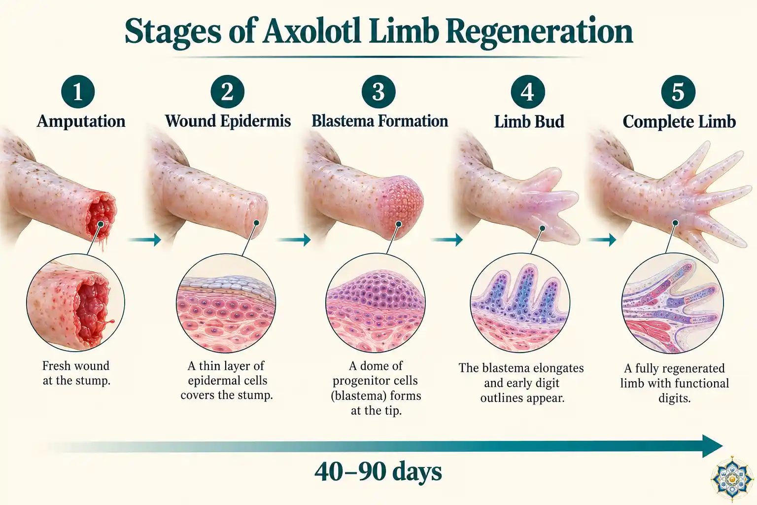
2. Step 1 — Wound Healing Without Scarring
Everything starts at the wound. And the very first thing the axolotl does differently from a human is also the most important: it doesn’t form a scar.
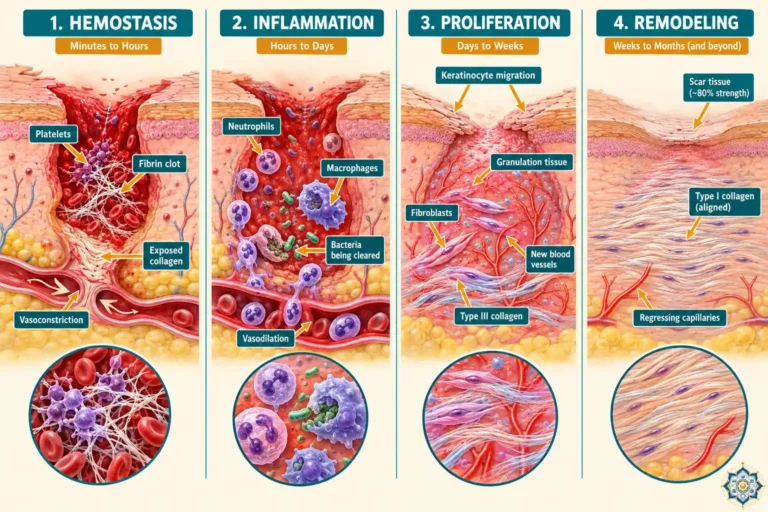
In humans, when tissue is damaged, the immune system launches an aggressive inflammatory response. Fibroblasts flood the wound site and deposit collagen rapidly, sealing the area with dense scar tissue. This is fast and effective — it prevents infection and bleeding — but it physically blocks any possibility of regeneration. The scar acts as a wall. (For the full four-phase sequence humans go through — hemostasis, inflammation, proliferation, and remodeling — see How Wounds Heal (And Why Scars Form).)
In the axolotl, something radically different happens:
- Hemostasis — Blood clotting stops the bleeding within minutes, as it does in mammals.
- Wound epidermis formation — Within 1–2 hours, epithelial cells from the surrounding skin migrate across the wound and form a thin, specialized layer called the wound epidermis (WE). This is not normal skin — it’s a signaling center.
- No fibrotic scarring — The axolotl suppresses the aggressive collagen deposition that creates scar tissue in mammals. TGF-β signaling, which drives fibrosis in humans, is modulated differently.
The wound epidermis is critical. If you experimentally remove it or replace it with a skin graft (which has a dermis), regeneration fails completely. The wound epidermis doesn’t just cover the wound — it actively sends molecular signals that trigger the next phase.
The wound epidermis as a signaling center
Research using single-cell RNA sequencing has revealed that the wound epidermis has three distinct layers — apical, intermediate, and basal — each with a different gene expression profile. The basal layer, sitting directly above the wound, acts as a progenitor reservoir. It expresses genes like krt17 and frem2 that are found only during regeneration — not in normal, intact skin.
The wound epidermis produces FGF (fibroblast growth factor) and EGF (epidermal growth factor), which signal the underlying tissue to begin the next step: dedifferentiation.
3. Step 2 — Dedifferentiation: Cells Going Backward
This is where axolotl regeneration enters territory that sounds like science fiction.
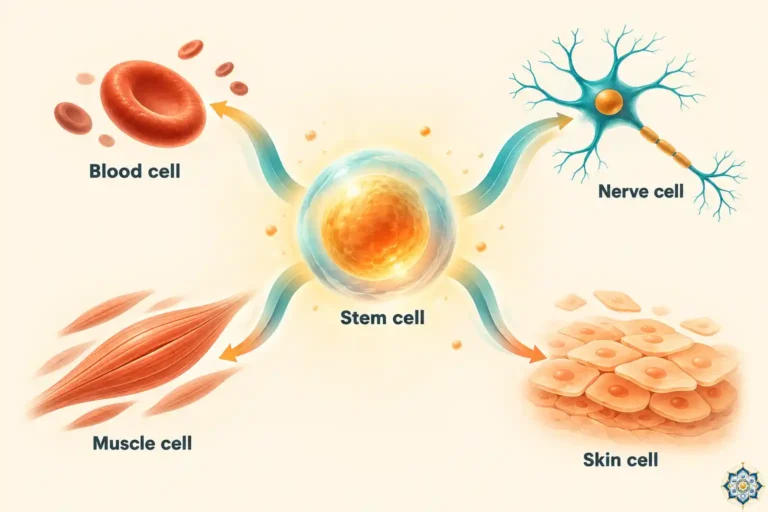
In your body, cell differentiation is a one-way street. A stem cell becomes a muscle cell, and that muscle cell stays a muscle cell for the rest of its life. It will never go back. In developmental biology, we say differentiation is terminal.
The axolotl breaks this rule.
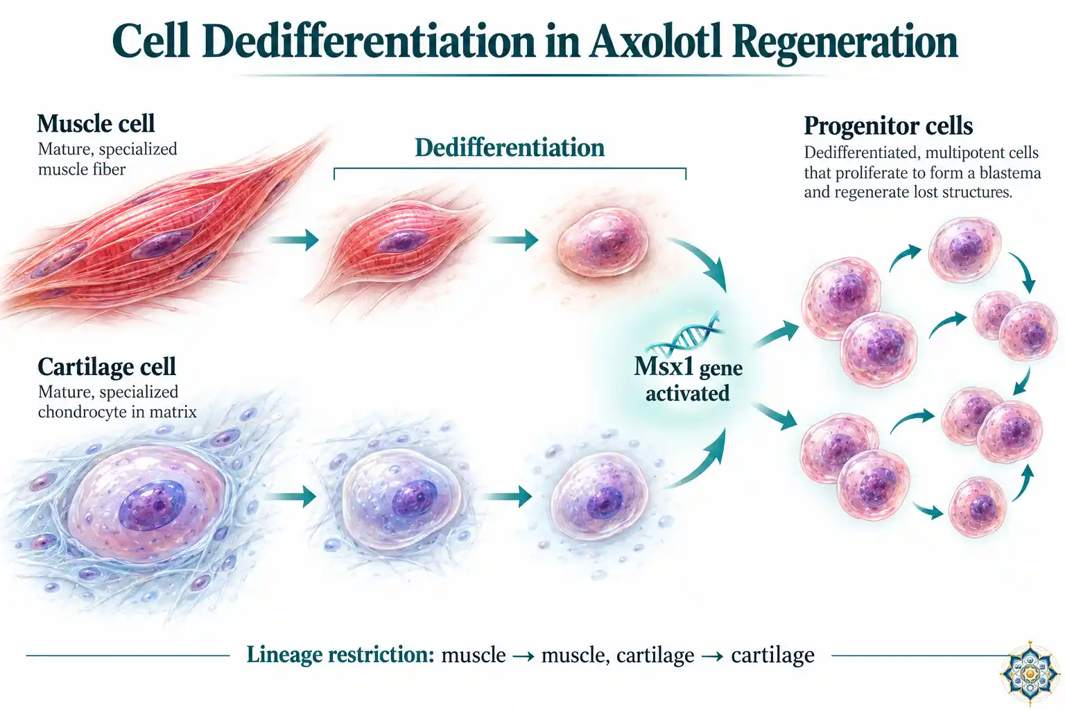
When the wound epidermis sends its signals, mature, specialized cells near the amputation site begin to dedifferentiate — they reverse their specialization. A muscle fiber that was contracting yesterday loses its contractile proteins, shrinks, re-enters the cell cycle, and starts dividing again. A cartilage cell that was embedded in solid matrix dissolves its surroundings and becomes mobile.
The Msx1 master switch
One of the key molecular players in dedifferentiation is Msx1, a transcription factor that promotes the reversal of cell identity. When Msx1 is experimentally activated in mouse muscle cells in a lab dish, those cells begin to lose their specialized features — hinting that the machinery for dedifferentiation isn’t unique to axolotls. Mammals have the gene. They just don’t turn it on at wound sites.
Lineage restriction — not a free-for-all
Here’s a crucial nuance: dedifferentiated axolotl cells don’t become truly pluripotent like embryonic stem cells. They retain lineage restriction — a memory of what tissue type they came from. Muscle cells that dedifferentiate will re-differentiate into new muscle. Cartilage cells will make new cartilage. Schwann cells (nerve-supporting cells) will remake Schwann cells.
This was demonstrated elegantly by grafting GFP-labeled (fluorescent) tissue into an axolotl before amputation. When the limb regenerated, researchers could track exactly which cell type produced which new tissue. The boundaries were clear: each lineage rebuilt itself.
Why lineage restriction matters: If dedifferentiated cells became fully pluripotent — able to become anything — the risk of uncontrolled growth (tumors) would be enormous. By keeping cells within their original lineage, the axolotl gets the benefits of cellular reprogramming without the cancer risk. This balance between plasticity and control is one of the most fascinating aspects of axolotl regeneration.
4. Step 3 — The Blastema: Biology’s Construction Site
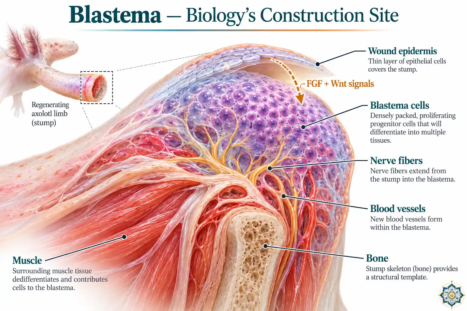
By about 7–14 days after amputation, the dedifferentiated cells have accumulated beneath the wound epidermis to form a visible dome of rapidly dividing cells. This structure is called the blastema — and it is the single most important structure in axolotl regeneration.
Think of the blastema as a biological construction site where the blueprint, the raw materials, and the workers are all cellular. It contains:
- Fibroblast-like blastema cells — the most abundant population, expressing genes like lum, dpt, and postn
- Myogenic progenitors — derived from Pax7+ satellite cells (muscle stem cells) and marked by Six1 and Eya1
- Schwann cell precursors — nerve-supporting cells expressing Sox2 and Foxd3
- Immune cells — macrophages, T cells, B cells, and dendritic cells
- Endothelial precursors — cells that will form new blood vessels
The blastema is not a disorganized blob. Single-cell RNA sequencing studies have revealed that it has a sophisticated internal architecture, with different cell populations occupying specific zones and expressing distinct sets of transcription factors.
Blastema growth signals
The blastema is maintained by a cocktail of growth signals:
| Signal | Source | Role in regeneration |
|---|---|---|
| FGF (fibroblast growth factor) | Wound epidermis | Promotes cell proliferation, prevents premature differentiation |
| Wnt | Multiple tissues | Maintains proliferative state, delays early differentiation |
| BMP (bone morphogenetic protein) | Blastema cells | Drives bone and cartilage formation during re-differentiation |
| Shh (sonic hedgehog) | Nerve-derived | Anterior-posterior patterning of digits |
| Retinoic acid | Proximal stump tissue | Encodes positional identity — determines which structures to rebuild |
| Neurotrophic factors (nAG) | Nerves + Schwann cells | Required for blastema cell proliferation — regeneration fails without them |
5. Step 4 — How Does the Axolotl Know What to Build?
This is the question that fascinates researchers most. When an axolotl loses a hand, it regrows a hand — not a foot, not a shoulder, not a random lump of tissue. The regenerate always matches what was lost, and it always builds from the cut point distally (outward). It never rebuilds tissue above the amputation.
How does the blastema know?
Positional memory
Cells in the axolotl limb carry positional information — molecular coordinates that encode where they sit along the limb axis. This is not a vague sense of location. It’s a precise molecular address encoded by the concentration of specific signaling molecules.
The primary position-encoding molecule is retinoic acid (a derivative of vitamin A). Retinoic acid exists in a gradient along the proximal-distal axis of the limb — highest near the body, lowest at the fingertips. When a limb is amputated, blastema cells read this gradient through retinoic acid receptors (RARs) and the enzyme CYP26B1 (which breaks down retinoic acid). A 2025 study published in Nature confirmed that retinoic acid breakdown by CYP26B1 is required for correct proximodistal identity during axolotl limb regeneration.
The transcription factor Hand2 also acts as a positional memory switch — it’s expressed differently along the limb axis and helps blastema cells remember whether they should build proximal (upper arm) or distal (finger) structures.
The classic experiment: If you treat an amputated wrist stump with extra retinoic acid, the blastema doesn’t regrow a hand — it regrows an entire arm, shoulder to fingertips. The retinoic acid “resets” the positional memory to a more proximal identity, so the blastema thinks it needs to rebuild everything from the shoulder down. This experiment, first performed in the 1980s, remains one of the most dramatic demonstrations of positional memory in biology.
The rule of distal transformation
Axolotl regeneration follows a strict rule: it only builds distally. If you amputate at the elbow, the blastema rebuilds the forearm, wrist, and hand — everything distal to the cut. It never rebuilds the upper arm. This tells us that the blastema reads its starting position and then executes a developmental program that runs from that point outward.
6. Step 5 — Rebuilding Tissue by Tissue
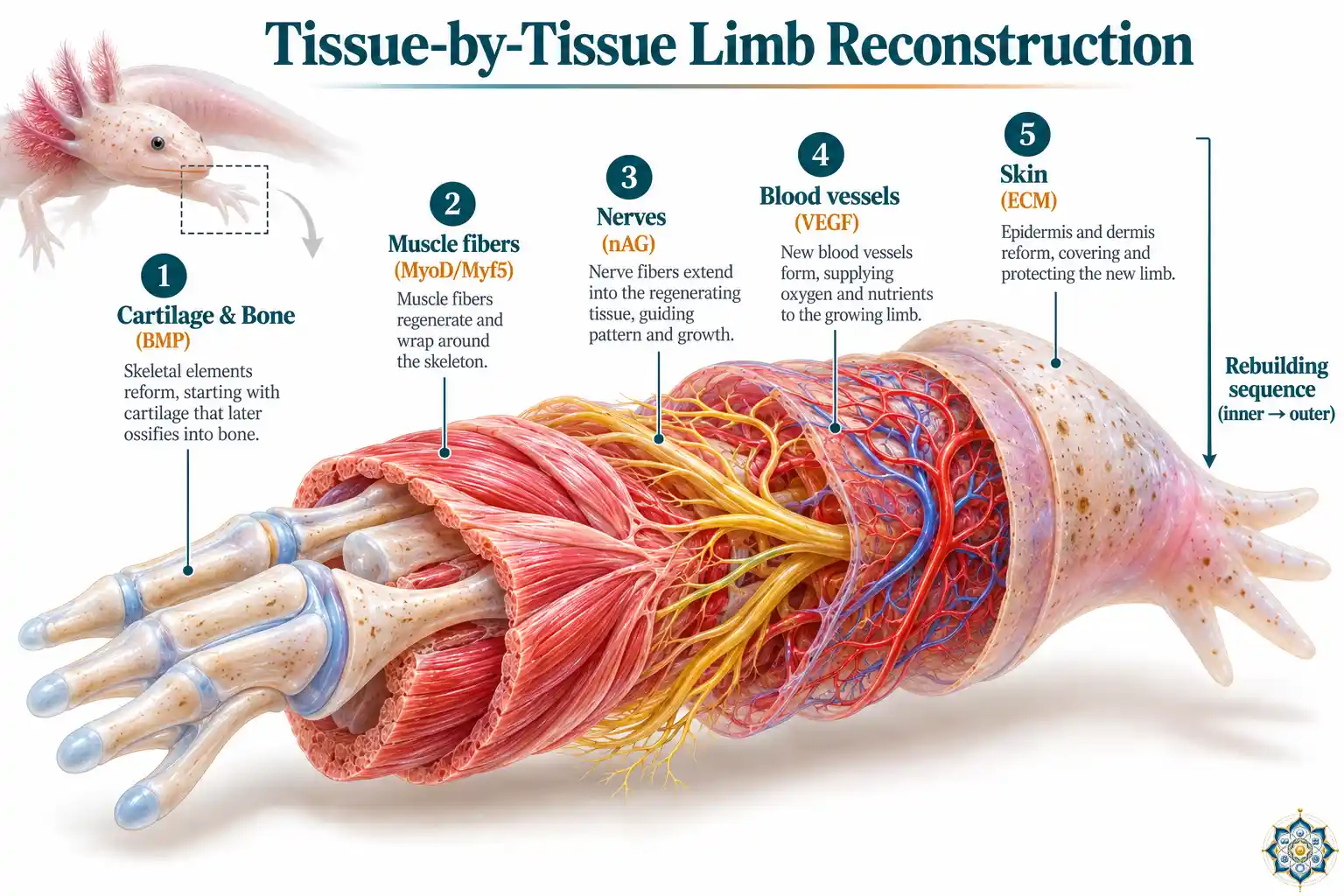
Once the blastema has reached sufficient size and its positional identity is established, cells begin to re-differentiate — converting back into specialized tissues. This happens in a specific sequence that mirrors embryonic limb development:
- Cartilage and bone — The skeletal elements form first, driven by BMP signaling. Cartilage condenses into the shapes of future bones.
- Muscle — Myogenic progenitors (marked by Myf5 and MyoD) begin fusing into muscle fibers, attaching to the forming skeleton.
- Nerves — Axons extend from the stump into the regenerate, guided by neurotrophic factors. Schwann cells remyelinate the growing nerves.
- Blood vessels — VEGF (vascular endothelial growth factor) drives the formation of new arteries, veins, and capillaries.
- Connective tissue and skin — Fibroblasts lay down the extracellular matrix. Skin closes over the regenerate — without scarring.
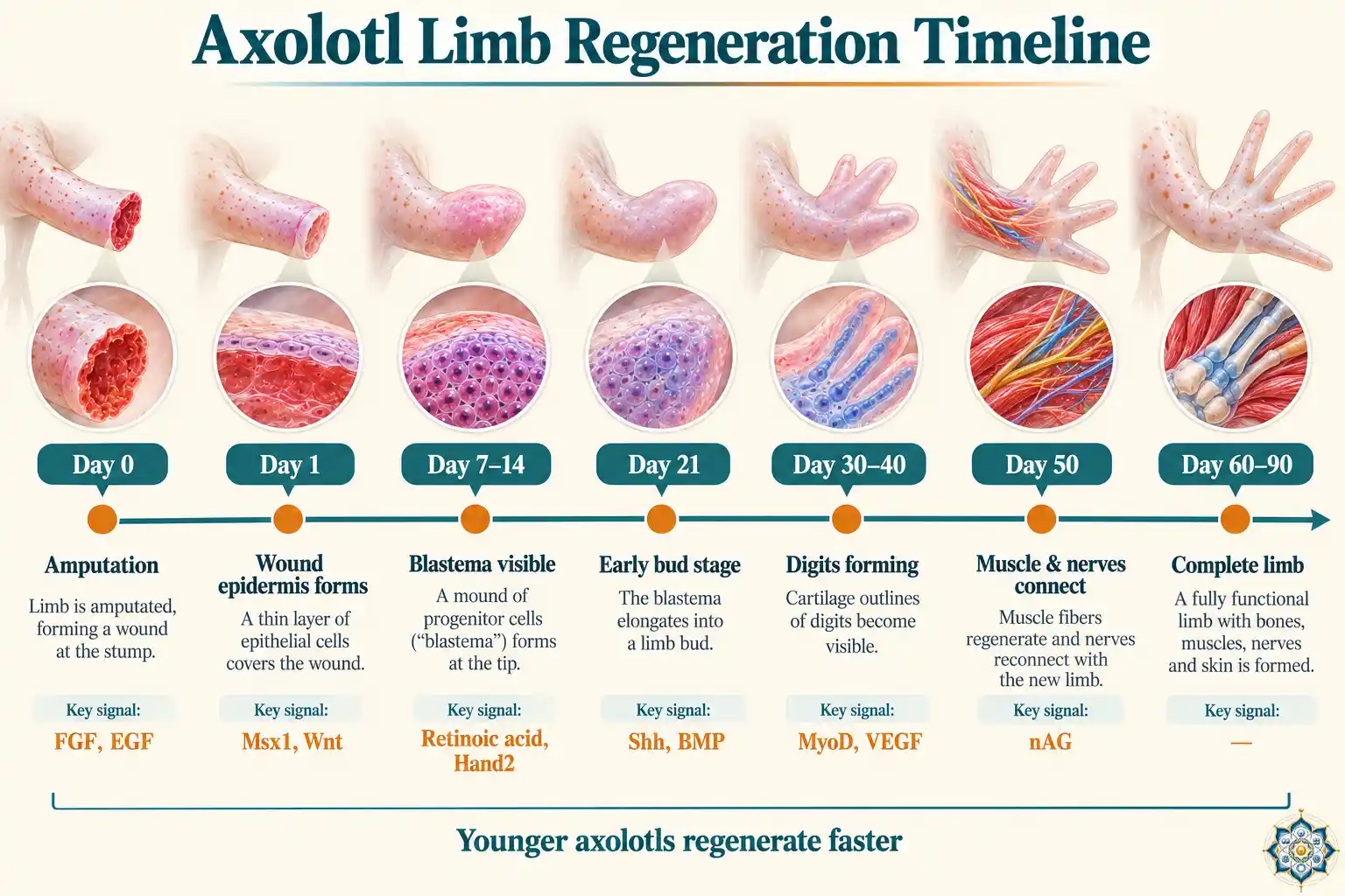
The entire process takes roughly 40–90 days, depending on the axolotl’s age, the location of the amputation, and environmental conditions (temperature, water quality). Younger axolotls regenerate faster. Distal amputations (closer to the fingertips) regenerate faster than proximal ones (closer to the body).
7. The Nerve Dependency — No Nerves, No Regeneration
One of the most striking discoveries in axolotl regeneration is that nerves are absolutely required. If you sever the nerves supplying a limb before amputation, the blastema fails to form. The wound closes, the epidermis covers it, dedifferentiation begins — but the cells never accumulate into a proper blastema, and regeneration stalls.
This nerve dependency was first demonstrated by Todd in 1823 and has been confirmed repeatedly since. The key molecule appears to be nAG (newt anterior gradient), a secreted protein produced by Schwann cells and the wound epidermis in response to nerve signals. When nAG is experimentally supplied to a denervated limb, regeneration is partially rescued — the blastema forms even without nerves.
This tells us something profound: the axolotl’s regenerative program isn’t just a property of the cells at the wound site. It requires active, ongoing communication between the nervous system and the regenerating tissue. The nerves don’t just supply electrical signals — they supply trophic (growth-promoting) molecules that the blastema cells depend on.
8. The Macrophage Switch — Inflammation vs. Regeneration
Perhaps the most medically relevant discovery in axolotl regeneration research is the role of macrophages — immune cells that are present in both axolotls and humans but behave very differently.
In humans, macrophages at a wound site are predominantly pro-inflammatory (M1 type). They release cytokines that recruit more immune cells, amplify inflammation, and activate fibroblasts to deposit scar-forming collagen. This response is fast and protective, but it prevents regeneration.
In axolotls, macrophages shift rapidly to a pro-regenerative profile. They clear debris efficiently, release anti-inflammatory signals, and provide growth factors that support blastema formation. When researchers experimentally depleted macrophages from axolotls before amputation, the result was dramatic: instead of regenerating, the limb formed permanent scar tissue — just like a mammal.
Godwin et al. (2013) used clodronate liposomes to remove macrophages from axolotls before limb amputation. Without macrophages, axolotls failed to form a blastema and instead developed fibrotic scar tissue. When macrophages were allowed to return, regenerative ability was restored — but only if the scar tissue was first removed. Once a scar forms, even the axolotl can’t regenerate through it. This proved that macrophages are not just helpful for regeneration — they are essential, and the critical window is the first few days after injury.
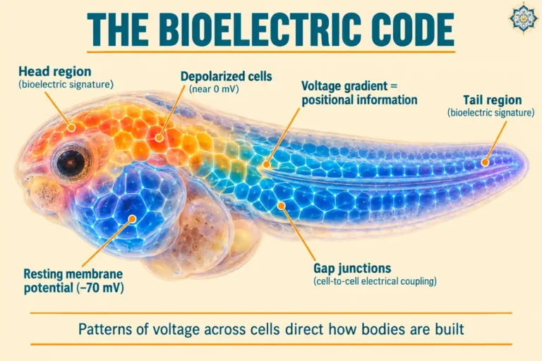
This finding has enormous implications for human medicine. It suggests that the difference between scarring and regeneration might be reprogrammable — if we can shift human macrophage behavior from pro-inflammatory to pro-regenerative at the right time, we might unlock regenerative potential that already exists in our tissues. A parallel line of research by Michael Levin’s lab at Tufts suggests this reprogramming might also work at the bioelectric level — by directly altering the voltage patterns across cells at the wound site, which carry their own instructions about whether to regenerate or scar.
9. The Largest Genome Ever Sequenced
In 2018, an international team led by researchers at the IMP in Vienna published the complete axolotl genome in Nature. It was a landmark achievement — and a massive challenge, because the axolotl genome is enormous.
| Species | Genome size (billion base pairs) | Estimated protein-coding genes |
|---|---|---|
| Axolotl | 32 | ~23,000 |
| Human | 3.2 | ~20,000 |
| Mouse | 2.7 | ~22,000 |
| Zebrafish | 1.4 | ~25,000 |
| Fruit fly | 0.14 | ~14,000 |
The axolotl genome is 10 times larger than the human genome — the biggest genome ever sequenced at the time. But here’s the surprise: it has roughly the same number of protein-coding genes as humans (about 23,000). The size difference comes almost entirely from massively expanded introns (non-coding DNA within genes) and transposable elements (mobile DNA sequences that have copied themselves throughout the genome).
This means the axolotl’s regenerative ability isn’t due to having thousands of special regeneration genes that humans lack. The functional genes are largely conserved. What differs is how those genes are regulated — when they turn on, where they’re expressed, and how strongly. This fits exactly with the central lesson of evo-devo: diversity across species, and across capabilities within a species, comes from how the ancient toolkit genes are deployed — not from inventing new ones.
The genome project also identified a few genes that are uniquely expressed in regenerating tissue — including some with no known function in any other species. These are being actively studied as potential keys to the regeneration puzzle.
10. Neoteny — The Salamander That Never Grows Up
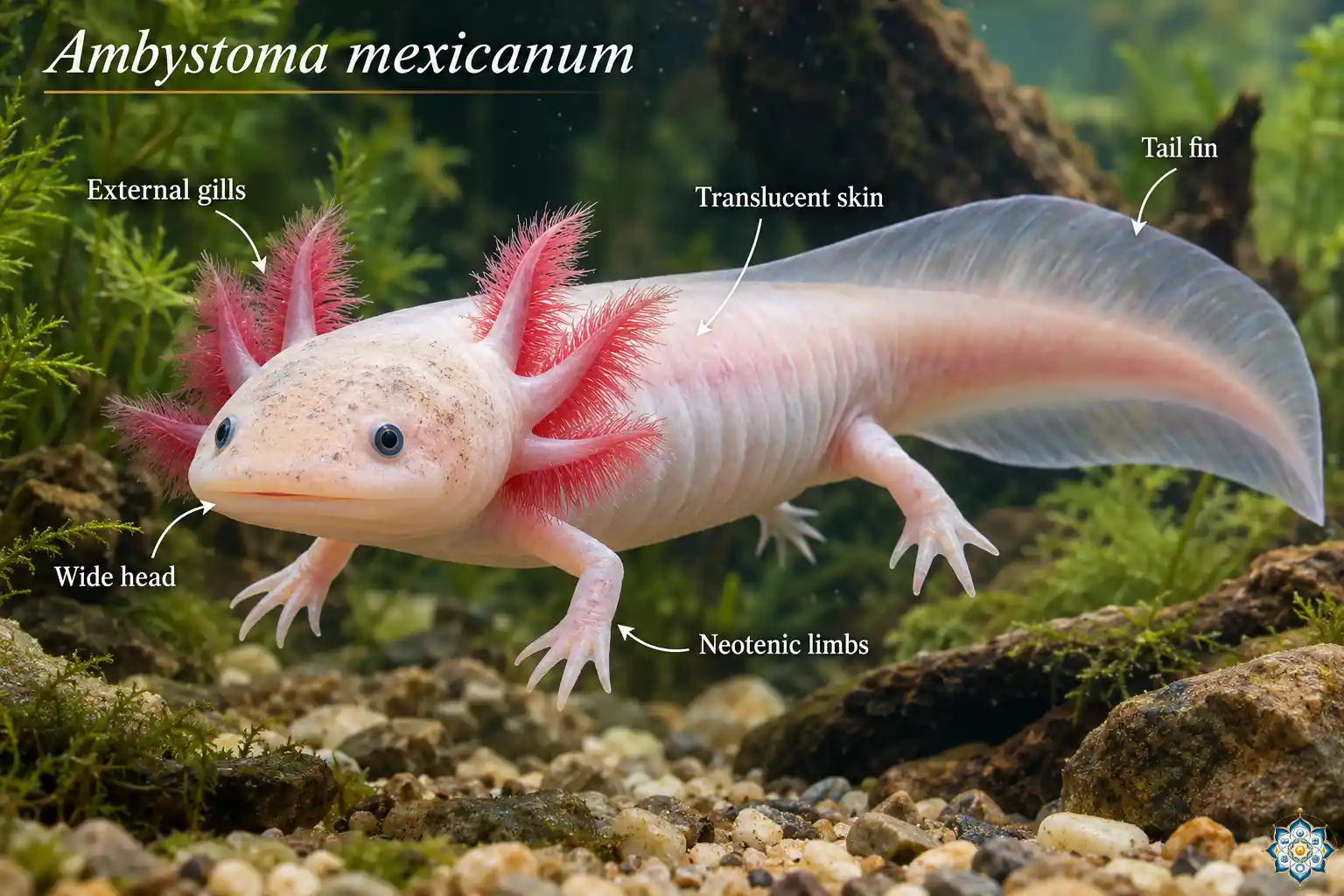
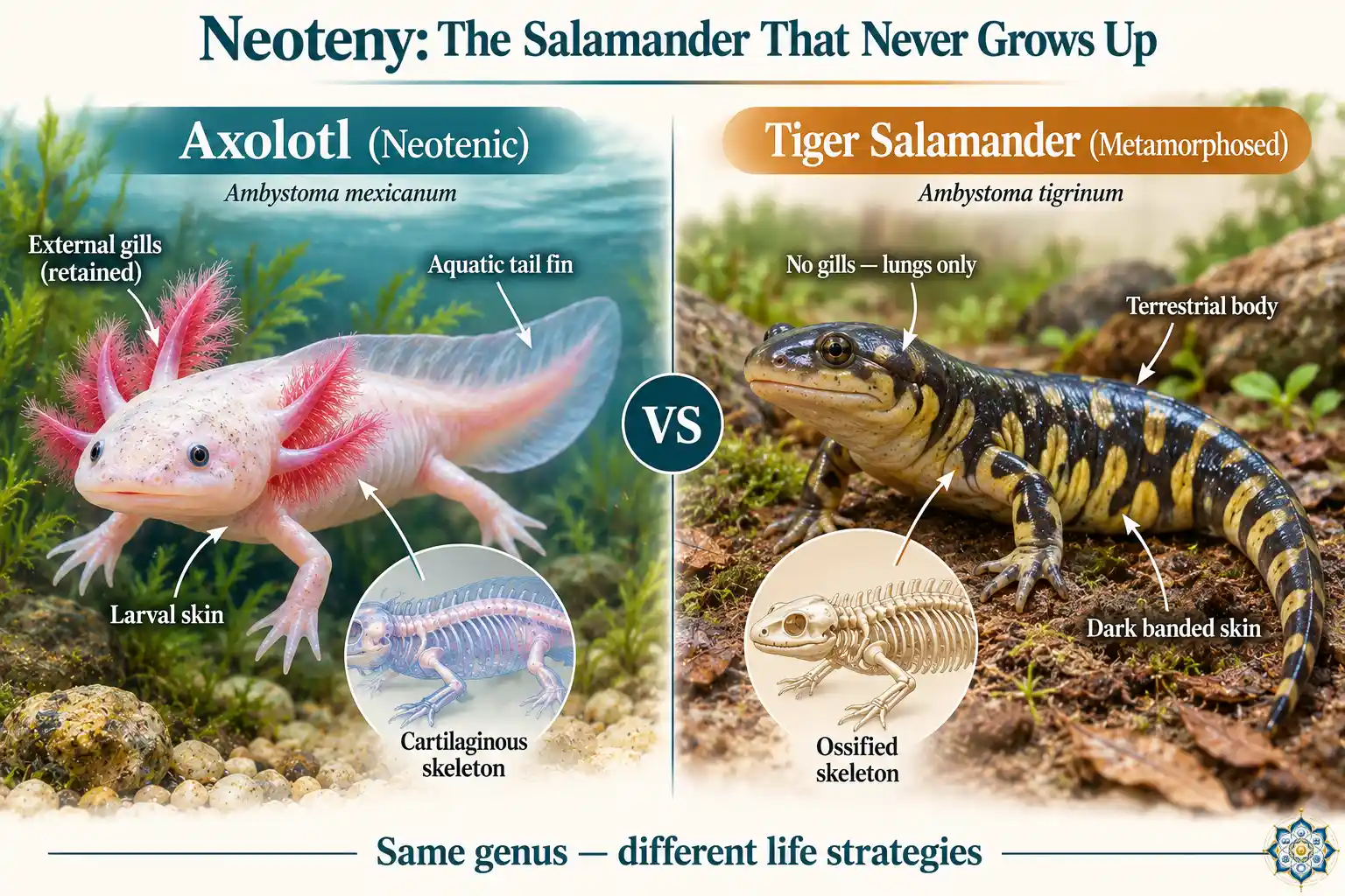
The axolotl is famous for another biological oddity: neoteny. Unlike most amphibians, the axolotl never undergoes metamorphosis. It retains its larval features — external feathery gills, a fin-like tail, cartilaginous (not ossified) skeleton, and aquatic lifestyle — throughout its entire adult life, even while reaching sexual maturity and reproducing.
Most salamanders lose their gills, develop lungs, and transition to a terrestrial life. The axolotl stays in the water forever, breathing through gills like an eternal juvenile. If you inject an axolotl with thyroid hormone (the signal that triggers metamorphosis in other amphibians), it will undergo metamorphosis — losing its gills, developing eyelids, and transitioning to a land-dwelling form. But it never does this naturally.
Does neoteny explain regeneration?
The connection between neoteny and regenerative ability is one of the most debated questions in the field. Several lines of evidence suggest a link:
- Larval tissues are more plastic. Embryonic and larval cells are inherently closer to the developmental programs needed for tissue construction. By staying in a larval-like state, the axolotl’s tissues may be “primed” for regeneration in a way that fully metamorphosed adult tissues are not.
- Metamorphosed salamanders regenerate more slowly. When axolotls are forced to metamorphose with thyroid hormone, their regenerative ability decreases — limbs regrow more slowly and with lower fidelity.
- Tadpoles lose regenerative ability at metamorphosis. Frog tadpoles can regenerate tails before metamorphosis but lose this ability as they develop into adult frogs — the transition correlates with changes in immune cell behavior and gene regulation.
However, the relationship isn’t simple. Some fully metamorphosed salamanders (like newts) retain significant regenerative ability, suggesting that neoteny alone isn’t the full explanation.
11. Why Humans Scar Instead of Regenerate
We’ve now seen the full axolotl playbook. So the natural question is: why can’t we do this?
The answer isn’t that we lack the genes. We have Msx1, Wnt, BMP, FGF, Hand2, Hox genes — nearly the entire axolotl toolkit. The problem is a tradeoff that evolution made in the mammalian lineage: speed vs. quality.
| Feature | Mammalian scarring | Axolotl regeneration |
|---|---|---|
| Speed | Days to weeks | Weeks to months |
| Infection risk | Low (rapid wound closure) | Higher (open wound longer) |
| Functional recovery | Partial — scar tissue is weaker | Complete — identical to original |
| Cancer risk | Lower (cells stay differentiated) | Potentially higher (cells re-enter cell cycle) |
| Immune response | Pro-inflammatory → fibrotic | Pro-regenerative → proliferative |
| Evolutionary advantage | Survive and move faster | Fully restore function over time |
For an axolotl sitting at the bottom of a lake, spending 60 days regrowing a leg is a viable strategy — it doesn’t need to run from predators. For an early mammal on the savanna, a 60-day open wound would be fatal. Evolution favored the fast, imperfect seal over the slow, perfect rebuild.
The specific blocks to regeneration in humans include:
- Aggressive fibrosis — Human fibroblasts rapidly deposit collagen, creating scar tissue that physically prevents blastema formation.
- Inflammatory macrophage dominance — Human macrophages sustain a pro-inflammatory state rather than shifting to the pro-regenerative profile seen in axolotls.
- Failure to dedifferentiate — Human somatic cells don’t naturally reverse their specialization. The genes are there, but the epigenetic locks preventing dedifferentiation are much stronger.
- Loss of nerve-derived growth signals — Mammalian nerves at wound sites don’t produce the trophic factors (like nAG) that axolotl nerves release.
But not all regeneration is lost. Humans can regenerate their liver (up to 70% of it), the tips of fingers (in children under ~7 years old, if the nail bed is intact), and some skin wounds in early fetal development heal without scarring. These exceptions suggest the ancient regenerative capacity isn’t entirely gone — it’s suppressed.
12. What Axolotl Regeneration Means for Human Medicine
Axolotl research isn’t just biology for biology’s sake. The molecular insights are directly informing multiple areas of translational medicine:
Scar-free wound healing
If we can replicate the axolotl’s suppression of fibrosis at wound sites — even partially — it would transform the treatment of burns, surgical scars, and internal organ scarring (liver fibrosis, cardiac fibrosis after heart attack, pulmonary fibrosis). The macrophage reprogramming pathway is the most promising target.
Spinal cord injury
Axolotls can regenerate severed spinal cords with full functional recovery. Understanding how they bridge the gap — which growth factors guide axons across the injury, how glia support reconnection — could inform therapies for paraplegia and quadriplegia.
Heart regeneration
After a heart attack, human cardiac muscle is replaced by scar tissue that can’t contract. Axolotl heart tissue regenerates instead of scarring. Research on the BMP/Wnt balance in cardiac regeneration is being explored for therapies that could induce human cardiomyocyte proliferation.
Understanding cancer
Axolotls routinely allow cells to dedifferentiate and re-enter the cell cycle — a process that in humans would raise cancer risk. Yet axolotls have remarkably low cancer rates. Understanding how they allow controlled cell division without malignant transformation could reveal new approaches to cancer prevention. This connects to a bigger reframing in oncology: cancer is development gone wrong — the same embryonic programs the axolotl safely activates for regeneration are what spiral out of control in human tumors.
Timeline reality check: Full human limb regeneration is likely decades away, if it’s possible at all. But more modest applications — scar-free wound healing, improved nerve regeneration, cardiac repair — could reach clinical trials within 10–15 years. The biology of axolotl regeneration is providing the molecular targets that pharmaceutical research is now pursuing.
13. Critically Endangered — The Paradox of the Axolotl
Here’s the tragic irony: the most regenerative animal on Earth is disappearing.
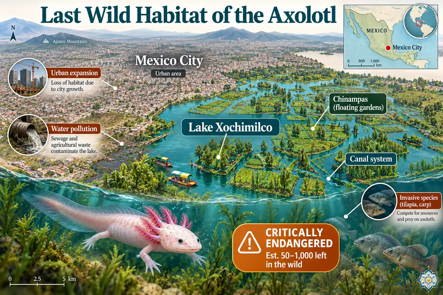
The axolotl exists in the wild in exactly one place — Lake Xochimilco and its connected canal system on the southern edge of Mexico City. The IUCN classifies the axolotl as Critically Endangered, with an estimated wild population of just 50 to 1,000 individuals.
The threats are multiple and severe:
- Habitat loss — Mexico City’s growth has drained and developed wetlands around the lake for centuries. The remaining canal system is a fraction of its historical extent.
- Water pollution — Agricultural runoff, sewage, and urban waste contaminate the remaining waterways.
- Invasive species — Non-native tilapia and carp, introduced for commercial fishing, eat axolotl eggs and juveniles and compete with adults for food.
- Water diversion — Water is diverted from the lake system for human use, shrinking the available habitat.
The paradox: while wild axolotls are vanishing, captive axolotls are thriving. Hundreds of thousands live in research laboratories worldwide (they’ve been a model organism since the 1860s), and millions more are kept as pets. The laboratory population descends from a colony brought to Paris in 1864 — just 34 individuals. This narrow genetic bottleneck means captive axolotls are highly inbred and may not represent the full genetic diversity of the wild population.
Conservation efforts centered on chinampas (traditional floating garden agriculture) in Xochimilco are attempting to restore axolotl habitat by cleaning water and creating predator-free refuges. But the challenges are enormous, and the clock is ticking.
14. Axolotl Limb Regeneration — Day-by-Day Timeline
| Time | Event | Key molecular players |
|---|---|---|
| 0–2 hours | Wound closes, blood clotting, wound epidermis begins migrating | FGF, EGF, fibrin clot factors |
| 6–12 hours | Wound epidermis covers the wound completely | Krt17, Frem2, TGF-β (suppressed) |
| Day 1–3 | Macrophages arrive — pro-regenerative profile, not pro-inflammatory | Trem2, ApoEB (debris clearance) |
| Day 3–7 | Dedifferentiation begins — mature cells revert to progenitor state | Msx1, FGF, Wnt |
| Day 7–14 | Blastema becomes visible — dome of dividing progenitor cells | Six1, Eya1, Pax7 (muscle), Sox2 (Schwann) |
| Day 14–21 | Early bud stage — rapid proliferation, positional memory is read | Retinoic acid, Hand2, Hox genes |
| Day 21–35 | Medium bud stage — patterning signals establish digit positions | Shh (anterior-posterior), BMP (skeletal) |
| Day 30–40 | Digits begin to form — cartilage condensation visible | BMP, Gdf5 (joint formation) |
| Day 35–50 | Muscle fibers form, nerves extend, blood vessels network | MyoD, Myf5, VEGF, nAG |
| Day 50–60 | Skin closes, pigmentation returns, limb approaches full size | Melanocyte migration, ECM remodeling |
| Day 60–90 | Internal refinement — bone ossification, tendon maturation, nerve myelination | Osteocalcin, collagen maturation |
15. Frequently Asked Questions
Can axolotls really regrow their limbs?
Yes. Axolotls can regenerate entire limbs — including bone, cartilage, muscle, nerves, and blood vessels — with full function restored. They can do this repeatedly throughout their lives. The regrown limb is virtually indistinguishable from the original.
What body parts can an axolotl regenerate?
Axolotls can regenerate all four limbs, the tail, portions of the brain, sections of the spinal cord, damaged heart tissue, parts of the lungs, the jaw, the skin (without scarring), portions of the eye (including the retina and lens), and the ovary. They are the most regenerative vertebrate known to science.
What is a blastema?
A blastema is a dome-shaped mass of rapidly dividing cells that forms at the site of amputation. It acts like a biological construction site — a reservoir of progenitor cells that will eventually re-differentiate into all the tissues needed to rebuild the missing structure.
How long does it take an axolotl to regrow a limb?
A full limb regeneration typically takes 40 to 90 days, depending on the axolotl’s age, the size of the amputated portion, and the water temperature. Younger axolotls regenerate faster. The blastema is visible within 7–14 days, and digits begin forming around day 30–40.
Why can’t humans regenerate limbs like axolotls?
Humans retain many of the same genes axolotls use for regeneration, but our wound-healing response favors rapid scarring over slow regeneration. When human tissue is damaged, fibroblasts quickly deposit collagen to seal the wound as scar tissue, which physically blocks the formation of a blastema. Axolotls suppress this scarring response.
Do axolotl cells turn back into stem cells during regeneration?
Not exactly. Mature cells near the wound undergo dedifferentiation — they revert to a progenitor-like state. But they retain lineage restriction — muscle cells give rise to new muscle, cartilage cells produce new cartilage. They don’t become fully pluripotent like embryonic stem cells.
How does the axolotl know which body part to regrow?
Blastema cells carry positional memory — molecular information encoded by gradients of retinoic acid and the activity of genes like Hand2 and Hox transcription factors. These signals tell the blastema whether to build a wrist or a shoulder, a finger or an upper arm.
Are axolotls endangered?
Yes. Axolotls are Critically Endangered according to the IUCN Red List. In the wild, they survive only in Lake Xochimilco near Mexico City, with an estimated population of 50 to 1,000 individuals. However, hundreds of thousands thrive in research laboratories and as pets worldwide.
What is neoteny and why does it matter?
Neoteny means retaining juvenile features into adulthood. Axolotls never undergo metamorphosis — they keep their larval gills, aquatic lifestyle, and cartilaginous skeleton. This permanent juvenile state may keep their cells closer to the developmental programs needed for regeneration.
Could axolotl research lead to human limb regeneration?
Possibly, though not soon. The most promising near-term applications are in scar-free wound healing, spinal cord repair, and heart tissue regeneration. Researchers have already identified key molecular switches that could be targeted therapeutically within the next 10–15 years.
16. References
- Nowoshilow, S. et al. (2018). The axolotl genome and the evolution of key tissue formation regulators. Nature, 554(7690), 50–55. doi:10.1038/nature25458
- Leigh, N. D. et al. (2018). Transcriptomic landscape of the blastema niche in regenerating adult axolotl limbs at single-cell resolution. Nature Communications, 9, 5153. doi:10.1038/s41467-018-07604-0
- Godwin, J. W., Pinto, A. R., & Rosenthal, N. A. (2013). Macrophages are required for adult salamander limb regeneration. Proceedings of the National Academy of Sciences, 110(23), 9415–9420.
- Kragl, M. et al. (2009). Cells keep a memory of their tissue origin during axolotl limb regeneration. Nature, 460, 60–65.
- McCusker, C. D. & Gardiner, D. M. (2013). Positional information is reprogrammed in blastema cells of the regenerating limb of the axolotl. PLoS ONE, 8(9), e77064.
- Del Moral-Morales, A. et al. (2024). Key proteins for regeneration in A. mexicanum. Scientifica, 2024, 5460694. doi:10.1155/2024/5460694
- Vieira, W. A. & McCusker, C. D. (2025). Retinoic acid breakdown is required for proximodistal positional identity during axolotl limb regeneration. Nature Communications, 16, 59497. doi:10.1038/s41467-025-59497-5
- Li, Y. et al. (2024). The genetic odyssey of axolotl regeneration: Insights and innovations. International Journal of Developmental Biology, 68, 103–116. doi:10.1387/ijdb.240111yl
- Kumar, A. & Brockes, J. P. (2012). Nerve dependence in tissue, organ, and appendage regeneration. Trends in Neurosciences, 35(11), 691–699.
- Whited Lab, Harvard University — Limb Regeneration Research. hscrb.harvard.edu
- IUCN Red List — Ambystoma mexicanum. Critically Endangered. iucnredlist.org
This article is written for educational purposes. The research findings and molecular mechanisms described here represent the current scientific understanding as of October 2026. Regenerative medicine applications discussed are in the research stage and are not currently available as clinical treatments. Content is reviewed against primary literature — see references above.
Follow HowLifeBuilds
@howlifebuilds