Why Cancer is Development Gone Wrong
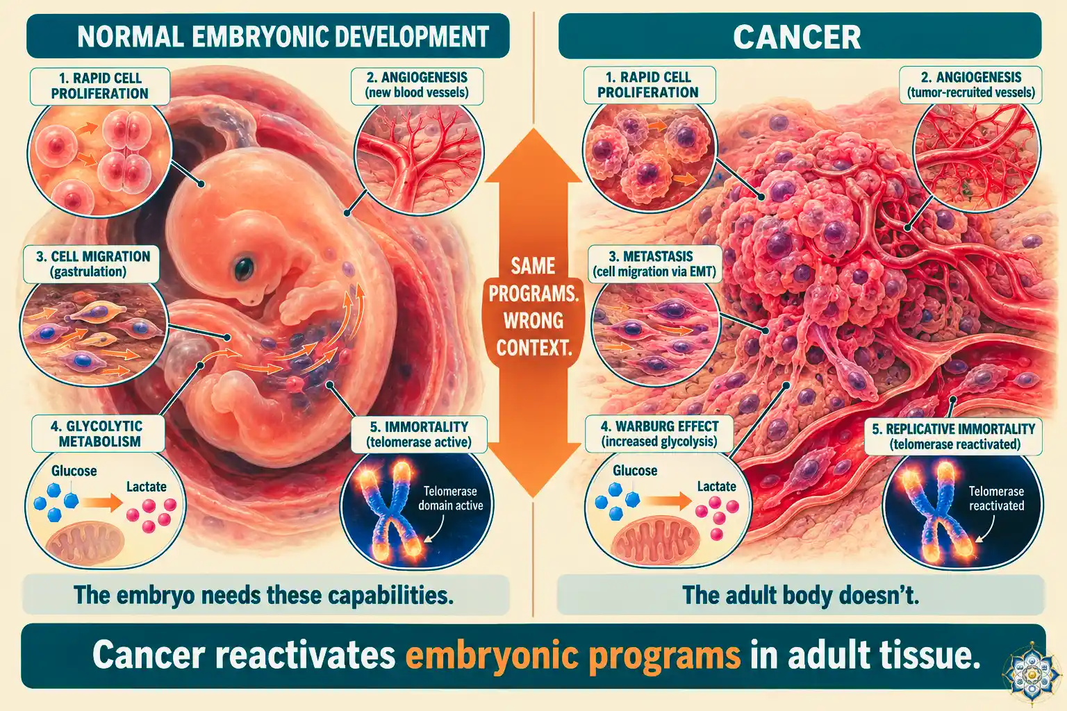
The idea that cancer is development gone wrong is one of the most important reframings in modern biology. Cancer is usually described as a disease — a collection of errors, a failure. But look at what a tumor actually does, and a stranger picture emerges.
Cancer cells divide rapidly, like embryonic cells. They build their own blood supply, like the placenta. They migrate to distant tissues, like gastrulating cells. They switch to a sugar-burning metabolism, like a fetus. They become immortal, like germline cells. They evade the immune system, like a developing embryo evading the mother’s.
Every one of these behaviors is a normal developmental program. They built you, every cell of you, during the first nine months of your existence. Then they were supposed to shut down. In cancer, they switch back on.
The phrase cancer is development gone wrong isn’t a metaphor — it’s an increasingly accepted framework in modern cancer biology. The hallmarks of cancer, mapped out by Douglas Hanahan and Robert Weinberg in a landmark 2000 paper and updated repeatedly since, read almost exactly like a list of embryonic capabilities. The 2022 update made the connection explicit by adding “unlocking phenotypic plasticity” to the official list — the ability of cancer cells to revert to earlier, more flexible developmental states.
Understanding cancer through this developmental lens changes what we think it is, how we think it starts, and most importantly, how it might be treated — not by killing cancer cells, but by convincing them to grow up.
The framing shift: cancer as misplaced embryology
For most of modern medicine, cancer has been treated as a problem of damaged DNA. Mutations accumulate, especially in genes that control cell division, and eventually a cell divides when it shouldn’t. The treatment logic followed: kill the mutated cells with chemotherapy or radiation.
This picture is correct but incomplete. Yes, mutations initiate most cancers — but they don’t explain what cancer cells actually do.
Consider what makes a cell cancerous. It has to:
- Keep dividing when it should stop
- Resist signals telling it to die
- Avoid becoming a specialized, non-dividing adult cell
- Recruit blood vessels to feed itself
- Switch its metabolism to sustain rapid growth
- Migrate through tissue barriers
- Settle and establish in new locations
Now consider what an embryo has to do. The exact same list.
Cancer isn’t inventing new tricks. It’s using programs that your cells already know — programs they ran at full capacity during embryonic development, then switched off. Those programs are preserved in your DNA throughout life. When the right suppressors fail, they can turn back on.
The hallmarks of cancer — read as embryonic behaviors
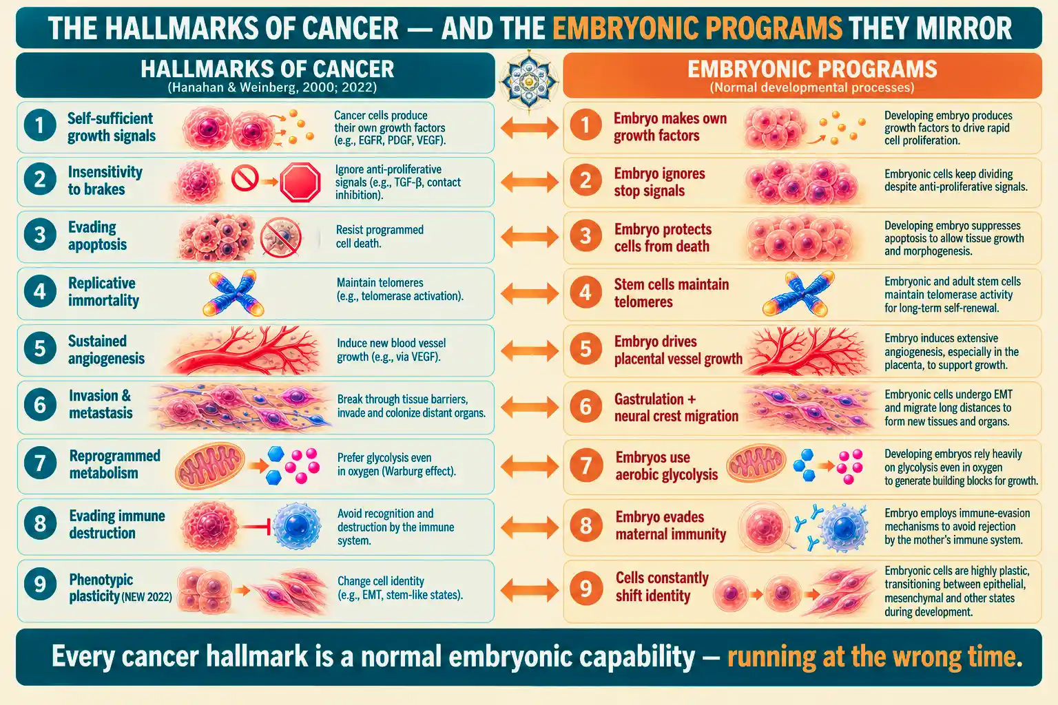
In 2000, Hanahan and Weinberg’s “Hallmarks of Cancer” paper argued that despite its complexity, cancer could be understood through a small number of acquired capabilities. The list has been updated twice — in 2011 and again in 2022 — and it reads like a developmental biology syllabus:
| Hallmark of cancer | The developmental program it borrows |
|---|---|
| Self-sufficient growth signals | Embryonic cells produce their own growth factors to drive rapid expansion |
| Insensitivity to growth brakes | Early embryo ignores most “stop dividing” signals until specific checkpoints |
| Evading apoptosis | Embryonic cells heavily resist programmed death to protect the developing body |
| Replicative immortality (telomerase) | Embryonic and stem cells maintain telomeres; adult cells do not |
| Sustained angiogenesis | Placenta and embryonic tissues drive aggressive new blood vessel growth |
| Tissue invasion and metastasis | Gastrulation and neural crest migration move cells across tissue boundaries |
| Reprogrammed energy metabolism (Warburg effect) | Embryonic cells use aerobic glycolysis — the same sugar-burning strategy |
| Evading immune destruction | Embryo evades maternal immune attack via immune privilege mechanisms |
| Phenotypic plasticity (new, 2022) | Developmental cells constantly shift identity, dedifferentiate, and transdifferentiate |
Every hallmark on the left has a mirror on the right. The developmental programs that built you are the same programs that cancer re-activates.
Phenotypic plasticity: the hallmark that puts development at the center
The 2022 update to the hallmarks added one that explicitly names the developmental connection: unlocking phenotypic plasticity. It describes three things cancer cells do that normal differentiated cells cannot:
1. Dedifferentiation
A specialized cell loses its mature identity and reverts to a progenitor-like state — the same mechanism axolotls use for regeneration. In axolotls, dedifferentiation produces a blastema that rebuilds a limb. In humans, uncontrolled dedifferentiation produces cancer stem cells.
2. Blocked differentiation
The cell starts to specialize but gets stuck partway. It never becomes a mature, non-dividing adult cell — it stays in a semi-embryonic, dividing state indefinitely. This is especially common in blood cancers like acute myeloid leukemia, where immature blast cells accumulate.
3. Transdifferentiation
A cell of one type converts to a cell of a different type — a developmental trick normally reserved for very specific contexts (like lens regeneration in some vertebrates). In cancer, it can mean a liver cell converting to behave like a bile duct cell, giving the tumor new properties mid-course.
All three of these are developmental mechanisms, running at the wrong time. And they explain one of the most frustrating facts in oncology: tumors are heterogeneous. A single tumor often contains multiple cell types, in different states of differentiation, with different drug responses. That heterogeneity isn’t noise — it’s a tumor using its developmental flexibility to find whichever cell state survives best under treatment.
Embryonic genes that turn back on in cancer
If cancer is development gone wrong, we should see embryonic genes reactivated in tumors. We do.
Pluripotency genes
The core genes that keep embryonic stem cells pluripotent — OCT4, SOX2, NANOG, and KLF4 — are the same four factors that Shinya Yamanaka used to reprogram adult cells into induced pluripotent stem cells. These genes are tightly silenced in adult tissue. In cancers of the breast, lung, prostate, and brain, they are reactivated — contributing to the stem-like, treatment-resistant properties of cancer cells.
Oncofetal antigens
Some proteins are produced only during fetal development and then shut off at birth. When cancer reactivates them, they can be detected in the bloodstream and used as diagnostic markers:
- Alpha-fetoprotein (AFP) — produced by fetal liver, reappears in liver cancer (hepatocellular carcinoma) and some germ cell tumors
- Carcinoembryonic antigen (CEA) — produced in fetal gut, reappears in colorectal and other cancers
- Human chorionic gonadotropin (hCG) — produced by placenta, reappears in testicular and other cancers
These oncofetal markers aren’t quirks. They’re direct evidence that cancer cells reactivate the gene expression programs of fetal tissue.
Toolkit genes misfiring
The evo-devo toolkit genes that pattern the embryo — Hox genes, Pax genes, Wnt signaling, Sonic Hedgehog, BMPs — are frequently dysregulated in cancer. Sonic Hedgehog hyperactivation drives basal cell carcinoma, the most common cancer in humans. Wnt pathway mutations drive the vast majority of colorectal cancers. These aren’t random mutations — they target the master switches of development.
The Warburg effect: cancer uses embryonic metabolism
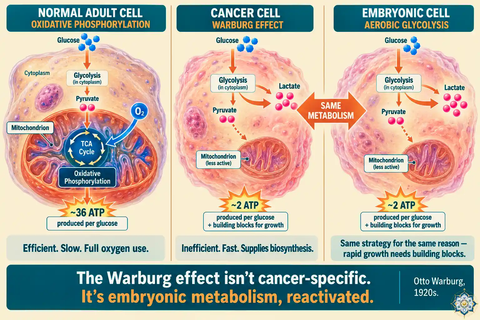
In the 1920s, German biochemist Otto Warburg noticed something strange about cancer cells. They burned sugar inefficiently. Instead of using oxygen to extract maximum energy from glucose (as normal adult cells do), they produced lactate — the same byproduct your muscles produce during intense exercise. They did this even when oxygen was abundant.
This is called the Warburg effect, or aerobic glycolysis. It’s wasteful of energy, but it’s fast — and it generates the molecular building blocks (amino acids, nucleotides, lipids) that rapidly dividing cells need.
For decades the Warburg effect was treated as a bizarre quirk. We now know it isn’t a quirk at all — it’s the normal metabolism of embryonic and fetal cells. Rapidly dividing embryonic tissue uses the same glycolytic strategy for the same reason: fast throughput and biosynthetic building blocks matter more than energy efficiency.
Cancer isn’t doing something unnatural. It’s doing something developmental.
EMT: how cancer borrows gastrulation to metastasize
The most dangerous thing cancer cells do is metastasize — leave the primary tumor and establish new tumors elsewhere in the body. About 90% of cancer deaths are caused by metastatic disease.
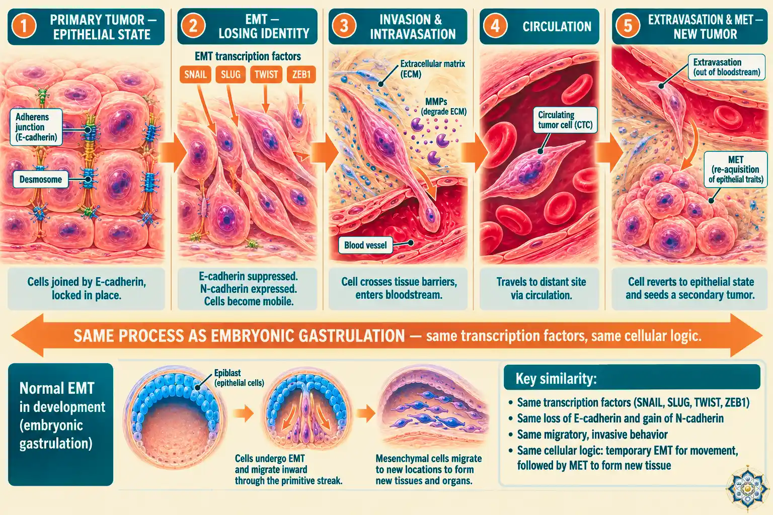
How do cancer cells, which started life stuck in a specific tissue, suddenly become mobile? They activate a developmental program called EMT (Epithelial-to-Mesenchymal Transition).
In EMT, a sheet-like, immobile epithelial cell loses its connections to its neighbors, dissolves its cell-cell adhesions, and becomes a free-moving mesenchymal cell. In an embryo, EMT is essential: it drives gastrulation (when the three germ layers form), neural crest migration (which produces much of the face, nervous system, and pigment cells), and heart valve formation. It’s one of the most important processes in development.
In cancer, EMT is hijacked to let tumor cells escape the primary site, invade blood vessels, travel through circulation, and settle in distant tissues — where they undergo the reverse process (MET, mesenchymal-to-epithelial transition) to establish a new tumor. The transcription factors that drive EMT in cancer — SNAIL, SLUG, TWIST, ZEB1 — are the exact same ones that drive EMT in the embryo.
Cancer stem cells: the developmental hierarchy inside a tumor
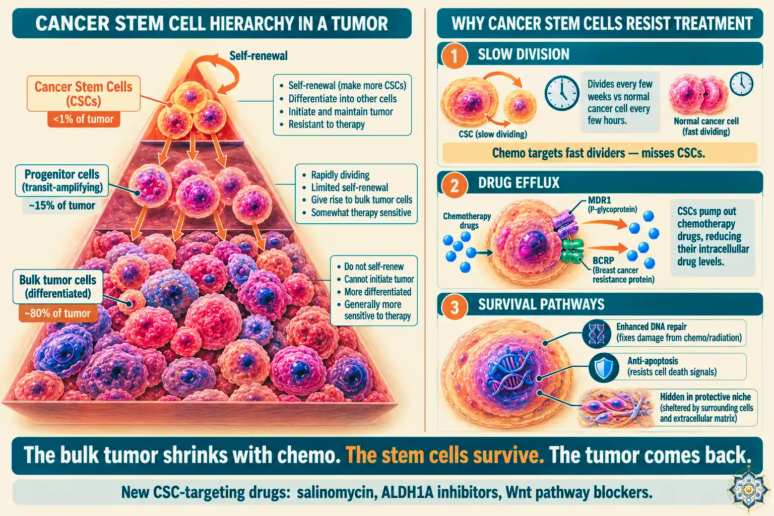
If cancer is development gone wrong, we’d expect tumors to be organized hierarchically — a small number of stem-like cells driving the growth, with their progeny making up the bulk of the tumor. This is exactly what research starting in the late 1990s has found.
In 1997, John Dick and Dominique Bonnet at the University of Toronto isolated the first cancer stem cells from acute myeloid leukemia. They found that a tiny subset of leukemia cells — fewer than 1 in 10,000 — could initiate the cancer when transplanted into mice. The rest of the leukemia cells, despite being malignant, couldn’t propagate the disease.
The term cancer stem cell (CSC) was formally proposed in 2001 by Reya, Morrison, Clarke, and Weissman. Since then, CSCs have been identified in brain, breast, colon, pancreatic, prostate, lung, and skin cancers. They share key properties with normal stem cells:
- Self-renewal — they can divide indefinitely while maintaining their stem-like state
- Multipotency — they can produce the different cell types found in the tumor
- Rare but influential — often less than 1% of the tumor, but responsible for its growth
Why cancer stem cells matter for treatment
Standard chemotherapy and radiation target rapidly dividing cells. Cancer stem cells often divide slowly, which lets them survive treatment. They also:
- Hide in protective niches within the tumor
- Pump drugs out using transporter proteins (MDR1, BCRP)
- Have enhanced DNA repair capacity
- Activate anti-apoptotic (survival) pathways
This is why many cancers come back after apparently successful treatment. The bulk tumor shrinks, but the cancer stem cells survive — and they’re all it takes to rebuild the tumor.
New approaches try to target CSCs specifically. In 2009, researchers found that salinomycin (originally used as a livestock antibiotic) killed breast cancer stem cells more than 100 times more effectively than paclitaxel chemotherapy. ALDH1A inhibitors and Wnt pathway blockers are being tested for similar reasons. The strategy is to shift from killing the fast-dividing cells to targeting the small reservoir of stem-like cells that regenerate the tumor.
The wound that never heals
In 1986, pathologist Harold Dvorak published a now-famous paper titled “Tumors: Wounds That Do Not Heal.” He noticed that the tissue environment inside a tumor looked remarkably like the environment of an actively healing wound: inflammation, blood vessel formation, extracellular matrix remodeling, immune cell infiltration, growth factor cascades.
The difference was that wounds eventually finish healing. Tumors don’t. They stay stuck in a perpetual “healing” state — recruiting fibroblasts, driving angiogenesis, remodeling matrix, suppressing the immune response — but never completing the process.
This framing matters because the same signals that drive wound healing drive cancer:
- TGF-β promotes healing early, promotes cancer invasion late
- VEGF builds new blood vessels in wounds and in tumors
- Macrophages clean up wounds and support tumor growth
- MMPs remodel matrix in healing and in metastasis
Treating cancer as a wound that won’t heal suggests new therapeutic targets — not killing cells, but completing the resolution phase that cancers can’t finish.
Why axolotls rarely get cancer
If cancer is development gone wrong, you might expect animals that are especially good at regeneration — like the axolotl, which can regrow entire limbs — to be especially prone to it. The opposite is true.
Axolotls have extraordinarily low cancer rates, even when exposed to carcinogens that reliably cause tumors in mice. Studies in which researchers deliberately injected axolotl tissue with chemicals that cause liver cancer in other animals produced no tumors.
Why? The leading theory connects back to the developmental framing. In axolotls, when cells gain the ability to proliferate aggressively — the first step toward cancer — they tend to channel that proliferation into regenerative rather than fibrotic/tumorigenic outcomes. Their macrophage behavior, bioelectric signaling, and intact developmental control systems appear to “finish the job” — producing new tissue rather than runaway growth.
Humans aren’t missing the regeneration genes. We’ve just locked them tightly behind programs that favor rapid scarring and tissue stability over regeneration. The flip side of that stability is that when control fails, the regenerative potential comes back as cancer instead of healing.
Bioelectricity, cancer, and the possibility of reversal
One of the most unexpected recent findings is that cancer cells have abnormal bioelectric signatures. In normal tissue, cells maintain specific resting membrane voltages that help coordinate their behavior with their neighbors. Cancer cells are typically more depolarized — their voltage pattern looks more like an embryonic cell than an adult cell.
Michael Levin’s lab at Tufts has demonstrated that this isn’t just a correlation. In frog experiments, inducing a depolarized bioelectric state in normal cells triggered tumor-like behavior. More remarkably, restoring normal bioelectric signaling — using light-activated ion channels — reversed tumors in some models. Cancer cells “rejoined” the surrounding tissue instead of being killed.
This fits perfectly with the developmental framing. If cancer is cells in an inappropriately embryonic state, correcting the voltage pattern that cues “which cell type to be” could, in principle, nudge them back toward normal adult identity.
Differentiation therapy: convincing cancer to grow up
If cancer is cells stuck in an embryonic state, the obvious treatment is to make them mature. This approach is called differentiation therapy, and it has already produced one of the most dramatic treatment successes in oncology.
Acute promyelocytic leukemia (APL) was once among the most lethal blood cancers. The disease is caused by immature white blood cells that get stuck mid-differentiation. In the 1980s, researchers discovered that treating APL patients with all-trans retinoic acid (ATRA) — a vitamin A derivative — pushed the stuck cells to continue differentiating into mature white blood cells, which then died naturally.
APL went from being nearly incurable to having a cure rate above 90%. The treatment doesn’t kill the cancer cells. It makes them grow up.
Expanding differentiation therapy to other cancers has proven difficult — each cancer type has its own developmental block, and finding the right signal to push past it is harder than it was for APL. But the proof of principle is powerful: in some cases, convincing cancer cells to complete development is more effective than destroying them.
Where mutations fit in
None of this means mutations don’t matter. They do — enormously. But the developmental framework changes how we think about which mutations matter and why.
Most cancer-driving mutations hit genes that control development itself. p53, mutated in more than half of all human cancers, is a master regulator that stops cells with damaged DNA from dividing. Rb controls when cells can enter the cell cycle. APC regulates the Wnt pathway. Myc integrates growth and proliferation signals. These aren’t random targets — they are nodes in the circuitry that controls developmental identity.
Alfred Knudson’s two-hit hypothesis (1971) explains why most cancers take decades to develop: a cell usually needs two independent mutations in a tumor suppressor gene before it loses function entirely. Most cancers require 4-8 driver mutations in such genes. That’s why cancer risk increases exponentially with age — the probability of accumulating enough driver mutations climbs with time.
The developmental lens doesn’t replace this picture. It completes it. Mutations provide the trigger. Development provides the program the triggered cells then run.
Key Takeaways
- Cancer isn’t a disease of random disorder — it’s cells reactivating embryonic developmental programs in adult tissue
- Every hallmark of cancer (proliferation, immortality, angiogenesis, metastasis, immune evasion, metabolic switching) is a mirror of a normal embryonic capability
- The 2022 addition of “phenotypic plasticity” to the hallmarks made the developmental connection official — cancer cells dedifferentiate, get stuck mid-differentiation, or transdifferentiate, just like embryonic cells
- Oncofetal antigens like AFP and CEA are direct evidence that cancer cells reactivate fetal gene expression
- The Warburg effect (cancer’s sugar metabolism) is the same metabolism used by rapidly dividing embryonic cells
- EMT, the developmental program that drives gastrulation, is hijacked by cancer for metastasis
- Cancer stem cells form a developmental hierarchy inside tumors — a small reservoir of stem-like cells drives the bulk growth
- Differentiation therapy (as in APL) proves that in some cases, convincing cancer cells to mature is more effective than killing them
Frequently Asked Questions
Is cancer caused by mutations or by developmental dysregulation?
Both — they’re not mutually exclusive. Mutations in genes like p53, Rb, APC, and Myc trigger cancer, but what makes a cell actually behave as cancer is the reactivation of embryonic programs those mutations unleash. Mutations are the ignition. Development is the engine that runs once ignited.
What are the hallmarks of cancer?
The hallmarks were defined by Hanahan and Weinberg in 2000 and updated in 2011 and 2022. The current list includes self-sufficient growth signals, insensitivity to growth brakes, evading apoptosis, replicative immortality, sustained angiogenesis, invasion and metastasis, reprogrammed metabolism, evading immune destruction, and phenotypic plasticity. Each one maps directly to a normal embryonic capability.
What are cancer stem cells?
Cancer stem cells are a small subset of cells within a tumor that can self-renew and produce the various cell types found in the tumor. They typically make up less than 1% of the tumor but drive its growth and resist standard treatment because they divide slowly and have enhanced survival mechanisms. They were first identified in acute myeloid leukemia by John Dick in 1997.
What is the Warburg effect?
The Warburg effect is cancer cells’ preference for aerobic glycolysis — producing energy by breaking glucose down to lactate even when oxygen is available. First described by Otto Warburg in the 1920s, it’s less efficient than normal mitochondrial respiration but faster, and it generates building blocks (amino acids, nucleotides, lipids) that rapidly dividing cells need. Embryonic cells use the same metabolic strategy.
Why do cancer cells become metastatic?
Metastasis occurs when cancer cells activate a developmental program called EMT (Epithelial-to-Mesenchymal Transition). EMT dissolves cell-to-cell adhesions and gives cells the ability to migrate — a program used in embryos during gastrulation and neural crest migration. The transcription factors (SNAIL, SLUG, TWIST, ZEB1) are the same ones used in development.
Why do axolotls rarely get cancer?
Axolotls have very low cancer rates even when exposed to strong carcinogens. The leading theory is that their intact regenerative control systems channel proliferative potential into regeneration rather than runaway tumor growth. Their macrophage behavior and bioelectric signaling appear to “complete the job” of tissue formation, preventing the sustained unresolved growth that characterizes tumors in mammals.
What is differentiation therapy?
Differentiation therapy treats cancer by forcing cancer cells to mature into normal, non-dividing adult cells rather than killing them. The classic success is acute promyelocytic leukemia (APL), which went from nearly incurable to over 90% cure rate using all-trans retinoic acid (ATRA), which pushes immature leukemia cells to complete differentiation into mature white blood cells.
Follow HowLifeBuilds
@howlifebuilds