How Wounds Heal (And Why Scars Form)
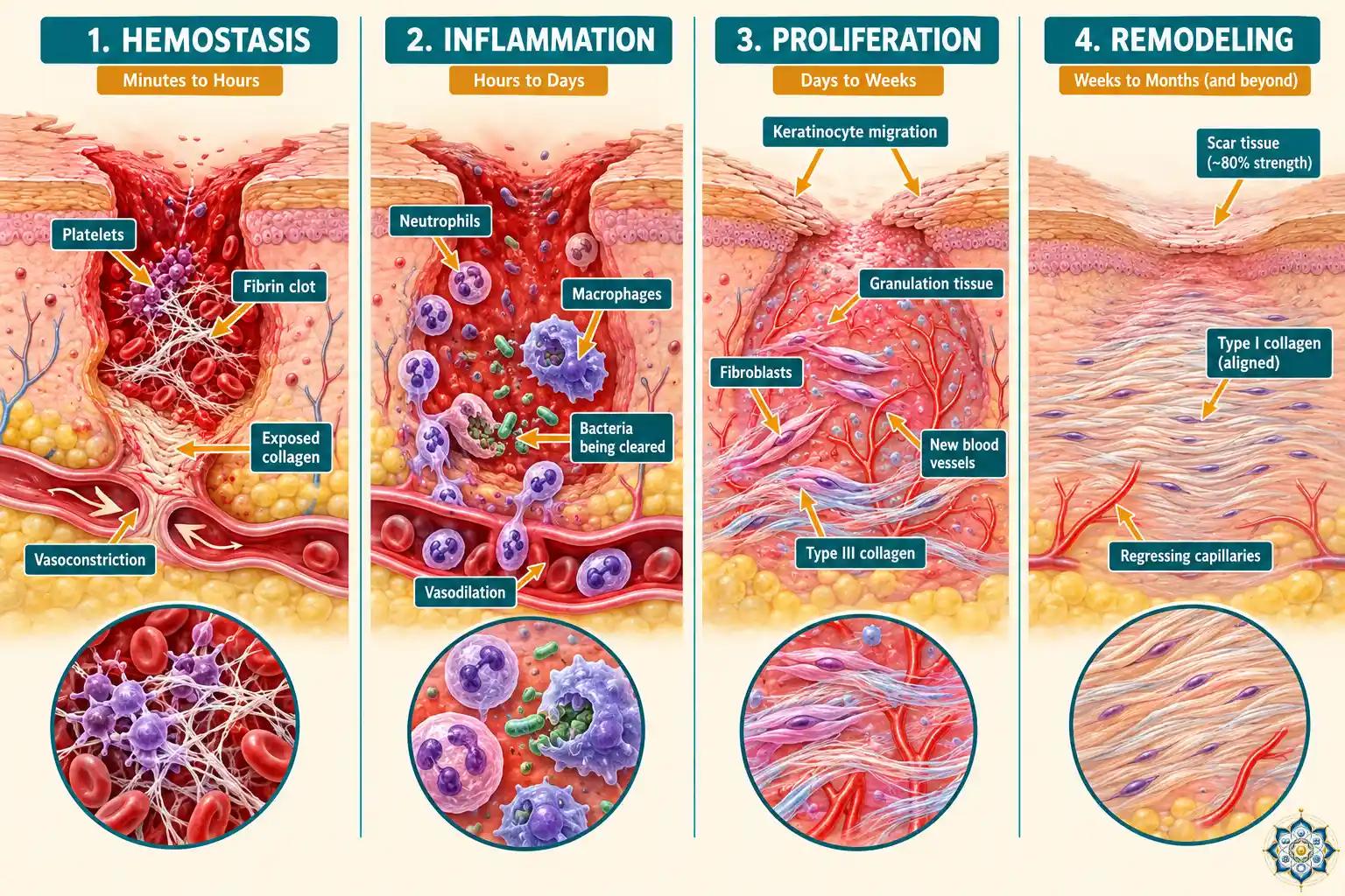
You cut your finger slicing onions. Within seconds, it bleeds. Within minutes, the bleeding slows. Within days, a scab forms. Within weeks, it peels away to reveal new skin — sometimes smooth, sometimes marked with a thin white line that stays for years.
This entire sequence looks simple from the outside. It’s anything but.
Wound healing is one of the most complex biological processes your body performs. It involves hundreds of cell types, dozens of molecular signals, and a choreography so precise that a failure at any single step can leave a wound open for months — or seal it with excessive scar tissue that never fully remodels.
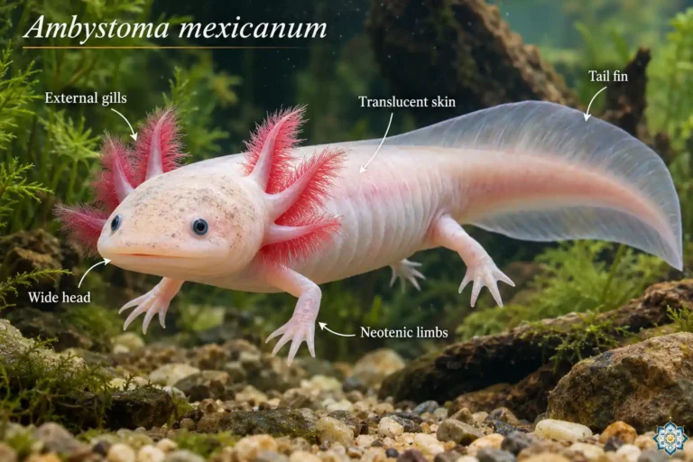
The strange part: your body could heal without scarring. Human fetuses do it in the womb. Axolotls do it their entire lives. But somewhere in our evolutionary history, we traded perfect regeneration for something faster and rougher — a system that prioritizes closing the wound quickly, even if the repair is imperfect.
This article walks through exactly what happens inside a wound, phase by phase, molecule by molecule — and why the result is a scar instead of perfect skin.
Phase 1: Hemostasis — stopping the blood (seconds to hours)
The instant skin breaks, the clock starts.
Damaged blood vessels release signaling molecules — thromboxane A₂ and endothelin — that cause immediate vasoconstriction. The vessels clamp down, reducing blood flow to the wound site. This buys time for the real repair to begin.
Within seconds, platelets rush to the scene. They stick to exposed collagen in the damaged tissue through a protein called von Willebrand factor, forming a plug. Then they activate — changing shape, clumping together, and releasing a cocktail of growth factors that will drive the entire healing process:
- PDGF (Platelet-Derived Growth Factor) — recruits fibroblasts, the cells that will later build new tissue
- TGF-β (Transforming Growth Factor-beta) — stimulates collagen production and modulates the immune response
- Serotonin — maintains vasoconstriction and increases vascular permeability
Simultaneously, the coagulation cascade fires. Thrombin converts a soluble blood protein called fibrinogen into fibrin — long, sticky threads that weave through the platelet plug to form a stable clot. This clot does three things at once: it stops bleeding, it seals the wound from bacteria, and it creates a temporary scaffold — a biological construction platform — for the cells that will arrive next.
Phase 2: Inflammation — cleaning the battlefield (hours to ~6 days)
If hemostasis is the emergency response, inflammation is the cleanup crew.
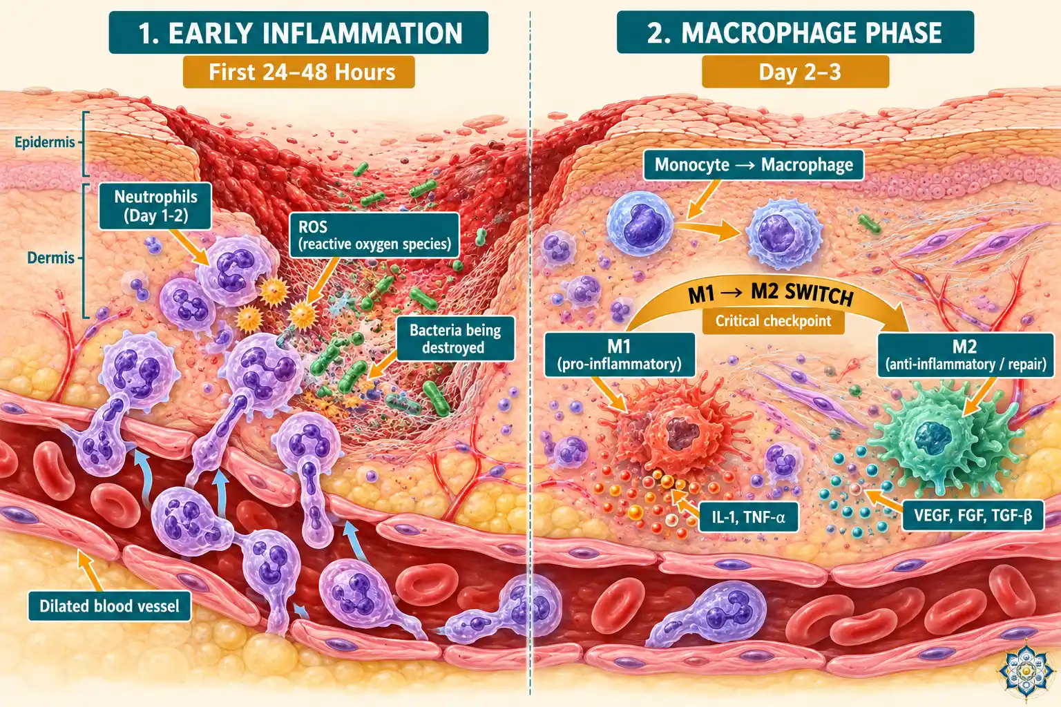
After vasoconstriction passes, the vessels do the opposite — they vasodilate. Histamine, prostaglandins, and complement fragments (C3a, C5a) flood the area, making blood vessels leaky and drawing immune cells in. This is why wounds swell, turn red, and feel warm. The inflammation isn’t the problem — it is the healing.
The first responders: neutrophils
Within hours, neutrophils — the most abundant white blood cells in your body — flood the wound. They dominate for the first 24 to 48 hours, doing two jobs: killing bacteria using reactive oxygen species (essentially molecular bleach) and breaking down damaged tissue using enzymes called matrix metalloproteinases (MMPs).
Neutrophils are blunt instruments. They destroy everything — bacteria, dead cells, even some healthy tissue in the crossfire. They’re effective, but messy. Within a few days, they self-destruct through a process called apoptosis (programmed cell death) and are cleared away.
The commanders: macrophages
Around day 2 to 3, monocytes arrive from the bloodstream and transform into macrophages — and these are the real directors of wound healing.
Macrophages come in two modes:
- M1 macrophages (pro-inflammatory) — continue the cleanup, secreting cytokines like IL-1 and TNF-α that maintain the inflammatory response and kill remaining pathogens
- M2 macrophages (anti-inflammatory) — shift the wound toward repair, releasing growth factors like VEGF, FGF, and TGF-β that signal fibroblasts and endothelial cells to begin building
This M1-to-M2 switch is one of the most critical checkpoints in wound healing. If it fails — if macrophages stay in M1 mode too long — the wound stalls in chronic inflammation and refuses to close. This is exactly what happens in diabetic foot ulcers and other non-healing wounds. It’s also central to why pathologist Harold Dvorak famously called cancer “the wound that never heals” — tumors sustain the same unresolved inflammatory and growth signals that healing tissue is supposed to shut down. (See Why Cancer is Development Gone Wrong for the full connection.)
Phase 3: Proliferation — building new tissue (day 4 to ~day 21)
Once the wound bed is clean, the construction begins.
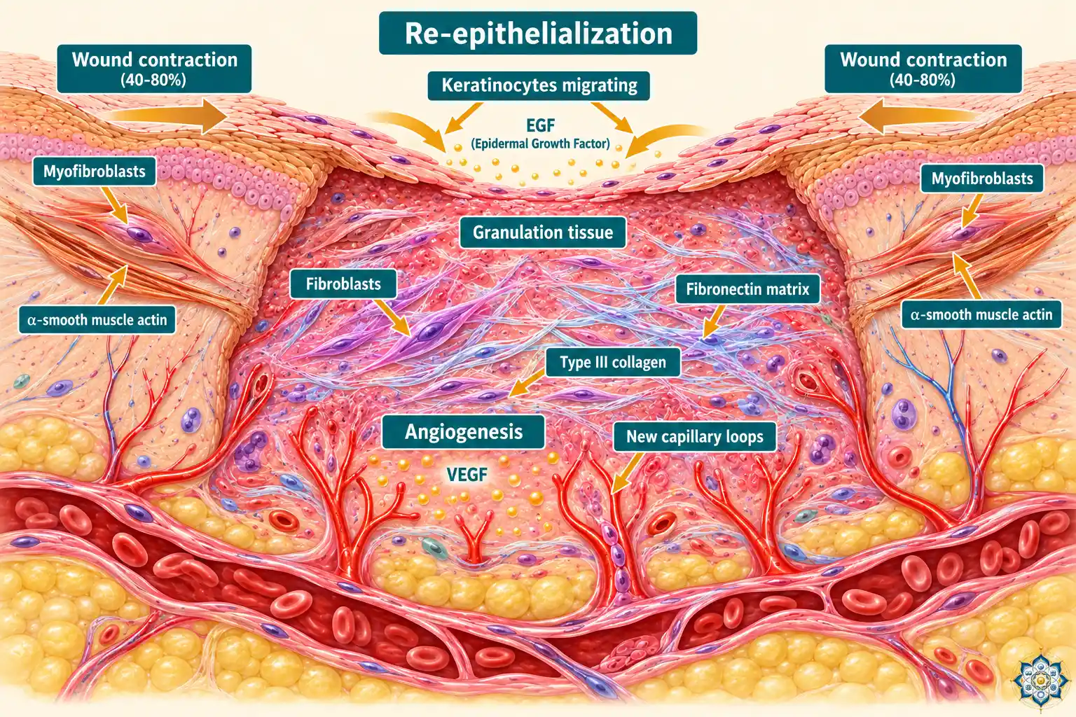
This is the most visually dramatic phase. Over two to three weeks, the wound fills with new tissue, new blood vessels, and new skin. Three processes happen simultaneously:
Angiogenesis: building new blood vessels
VEGF (Vascular Endothelial Growth Factor), released by M2 macrophages, drives endothelial cells to sprout new capillaries from existing vessels. These tiny loops push into the wound bed, delivering oxygen and nutrients to the construction site. This network of new capillaries gives healing tissue its characteristic red, granular appearance — which is why it’s called granulation tissue.
Fibroplasia: the collagen scaffold
Fibroblasts — the workhorses of wound healing — migrate along the fibrin scaffold left behind by the blood clot. They begin depositing type III collagen, along with fibronectin and proteoglycans, gradually building a new extracellular matrix.
Type III collagen is thinner and more flexible than the type I collagen found in mature skin. Think of it as temporary scaffolding — strong enough to hold the structure together, but not the final material. That replacement comes later, in the remodeling phase.
Re-epithelialization: restoring the surface
Keratinocytes at the wound edges lose their cell-to-cell connections and begin crawling across the granulation tissue like tiles being laid across a floor. They’re guided by EGF (Epidermal Growth Factor) from platelets and macrophages. When migrating keratinocytes from opposite edges meet, contact inhibition signals them to stop — the surface is sealed.
Wound contraction: pulling it together
Some fibroblasts transform into myofibroblasts — cells that express alpha-smooth muscle actin and can physically contract. They grip the wound margins and pull them together, reducing the open wound area by 40 to 80 percent. This is why a large scrape ends up much smaller than it originally was.
Phase 4: Remodeling — the long rebuild (day 21 to over 1 year)
The wound is closed. The surface looks healed. But beneath the skin, the repair continues for months — sometimes more than a year.
This phase is defined by one critical process: the collagen switch.
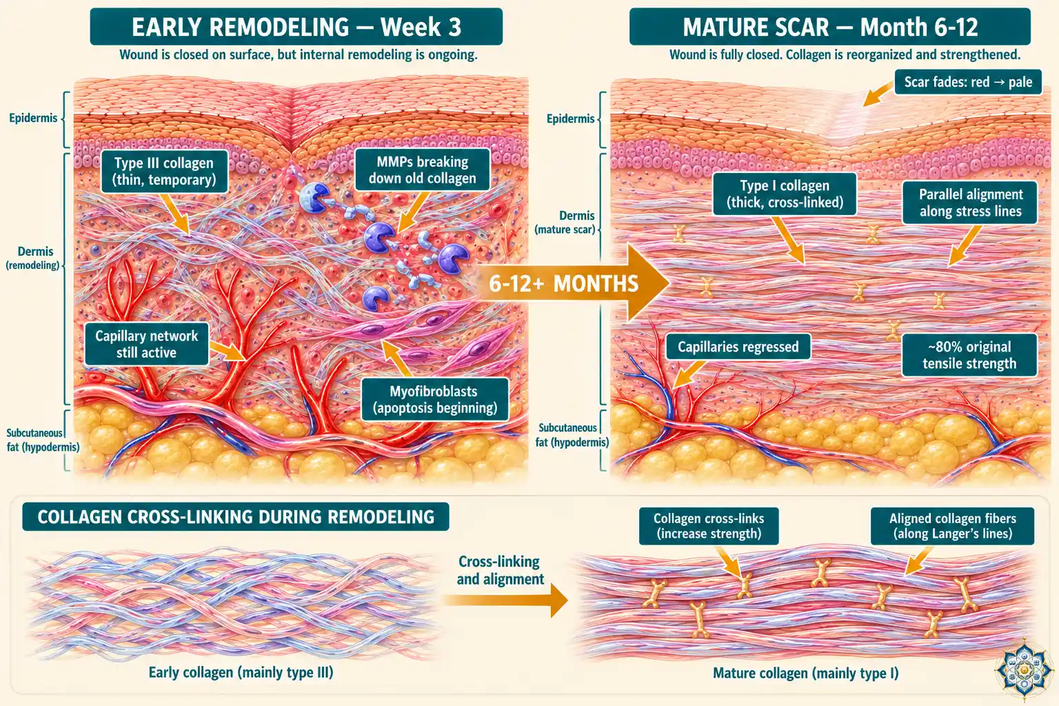
Type III collagen — the thin, temporary scaffolding laid down during proliferation — is gradually broken down by matrix metalloproteinases (MMPs) and replaced by type I collagen, which is thicker, stronger, and more cross-linked.
As new collagen fibers are deposited, they align along lines of mechanical stress — the directions your skin is regularly pulled or stretched. Excess blood vessels regress, myofibroblasts undergo apoptosis, and the metabolic activity of the wound slowly returns to normal.
The scar fades from red to pale as the capillary network shrinks.
But here is the critical limitation:
The molecular signals that drive wound healing
Every phase of wound healing is coordinated by a specific set of growth factors and cytokines. Here’s the complete signaling map:
| Signal | Source | Primary Role |
|---|---|---|
| PDGF | Platelets, macrophages | Recruits fibroblasts to the wound site |
| TGF-β | Platelets, macrophages, T cells | Drives collagen synthesis; modulates immune response |
| VEGF | Macrophages, keratinocytes | Stimulates new blood vessel growth (angiogenesis) |
| FGF | Macrophages, mast cells | Promotes fibroblast and endothelial cell proliferation |
| EGF | Platelets, macrophages | Drives keratinocyte migration (re-epithelialization) |
| TNF-α | M1 macrophages | Pro-inflammatory signaling; angiogenesis at low doses |
| IL-1 | M1 macrophages | Amplifies inflammatory response |
| MMPs | Neutrophils, macrophages, fibroblasts | Break down damaged matrix; remodel collagen during maturation |
Why scars form: the collagen problem
The answer to “why do scars look different from normal skin?” comes down to one thing: collagen organization.
In undamaged skin, collagen fibers are arranged in a complex, basket-weave pattern — fibers running in multiple directions, interlocking at various angles. This gives skin its remarkable combination of strength, flexibility, and elasticity.
In scar tissue, collagen fibers align in parallel bundles along a single axis — the direction of greatest mechanical tension. The result is a tissue that is structurally intact but fundamentally different: stiffer, less elastic, unable to stretch the way normal skin does. It has no hair follicles, no sweat glands, no sebaceous glands. It’s a patch, not a restoration.
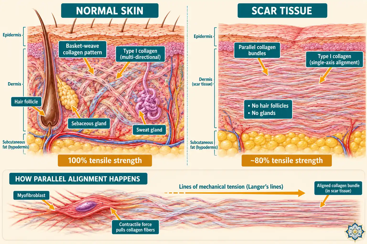
Why does collagen align this way?
It’s driven by mechanical tension.
During healing, fibroblasts become myofibroblasts — contractile cells that pull the wound closed. As they generate force, they align collagen fibers along the direction of that force. The collagen, in turn, guides more fibroblasts to deposit even more collagen in the same direction. It’s a feedback loop: tension drives alignment, alignment drives more tension.
Research from NJIT has shown that this process is governed by anisotropy — directional force in the matrix. Fibroblasts naturally align along Langer’s lines (the natural lines of tension in human skin), and when this alignment isn’t regulated, collagen builds up in thick, disorganized layers — producing raised or thickened scars.
The role of TGF-β
The growth factor TGF-β plays a central role in scar formation. It stimulates fibroblasts to produce collagen aggressively and promotes their transformation into myofibroblasts. In normal healing, TGF-β activity gradually decreases as the wound closes. But when TGF-β signaling stays elevated — or the ratio of its subtypes shifts — scarring becomes excessive.
This is where fetal healing provides a crucial insight.
Why fetuses heal without scars
In the first two trimesters of development, human fetuses heal skin wounds without any scarring at all. The wound simply regenerates — new skin, new hair follicles, new glands. Identical to the original.
By the third trimester, this ability disappears. The fetus begins scarring like an adult.
What changes? Several things:
- TGF-β ratio shifts: Fetal wounds have high levels of TGF-β3 (anti-fibrotic) and low levels of TGF-β1 (pro-fibrotic). Adult wounds are the opposite — high TGF-β1, low TGF-β3. This ratio shift is one of the strongest predictors of whether a wound will scar.
- Collagen composition: Fetal wounds produce a higher ratio of type III collagen to type I collagen, and the collagen is deposited in a reticular (net-like) pattern rather than parallel bundles.
- Reduced inflammation: Fetal wounds have significantly less inflammatory response — fewer neutrophils, fewer macrophages, lower levels of pro-inflammatory cytokines. The immune system hasn’t fully matured yet.
- Different extracellular matrix: The fetal matrix is rich in hyaluronic acid, which promotes cell migration and keeps the matrix hydrated and flexible, facilitating regeneration over fibrosis.
In other words, the default program for human wound healing is regeneration. Scarring is what happens when that program gets overridden by the adult immune system and its inflammatory cascade.
Why axolotls never scar — and what it means for humans
The axolotl doesn’t just heal wounds without scarring. It regenerates entire limbs, sections of its heart, spinal cord, and even parts of its brain. No scar tissue. Perfect reconstruction.
The key difference between axolotl healing and human healing comes down to macrophage behavior.
Research by James Godwin at the MDI Biological Laboratory found that axolotl macrophages promote regeneration rather than fibrosis. When Godwin’s team depleted macrophages from axolotls, something remarkable happened: the animals lost their regenerative ability and formed scars instead — just like humans.
This means scarring isn’t the default. It’s a specific outcome driven by how macrophages respond to injury. The axolotl’s macrophages send different signals than ours, steering the repair toward regeneration instead of fibrosis.
Godwin’s conclusion: “Once a scar has formed, it’s game over in terms of regeneration.” The scar physically blocks the regenerative process. If you could prevent the scar from forming in the first place — by making human macrophages behave more like axolotl macrophages — you might unlock the regenerative capacity that humans already carry in their genome but can’t access.
Not all scars are equal: the four types
The type of scar that forms depends on how the healing process unfolds — and what goes wrong during it:
1. Normal (flat) scars
The standard outcome. Initially red or pink, they flatten and fade over 6 to 18 months. The collagen remodeling phase completes normally, and the scar reaches about 80% of original skin strength.
2. Hypertrophic scars
Raised, thick, and red. They form when collagen production during the proliferation phase is excessive — too many fibroblasts, too much TGF-β, too much collagen deposited too quickly. They stay within the boundaries of the original wound and usually flatten over 1 to 2 years.
3. Keloid scars
Like hypertrophic scars, but worse. Keloids grow beyond the original wound boundaries, invading surrounding healthy skin. They’re caused by fibroblasts that don’t receive the apoptosis signals that normally stop collagen production. Keloids can continue growing for years and rarely resolve on their own.
4. Atrophic scars
Sunken or depressed scars — the opposite of keloids. They form when not enough collagen is produced during healing, leaving a pit or indentation. Acne scars and chickenpox scars are the most common examples.
Can we prevent scars? Where the research stands
The science of scar prevention is moving from “manage it after it forms” to “prevent it from forming in the first place.” Several promising approaches are in development:
Macrophage reprogramming
Building on the axolotl research, Godwin’s team is developing a topical hydrogel that would modulate human macrophage behavior at the wound site — essentially making them behave more like axolotl macrophages, promoting regeneration over fibrosis. This is still in the proposal stage, not yet a tested treatment.
Bioelectric reprogramming
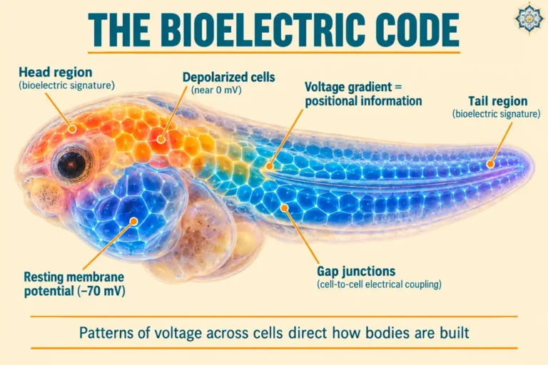
Michael Levin’s lab at Tufts has shown that voltage patterns across cells carry their own instructions about regeneration versus scarring. His team’s BioDome device creates a controlled electrochemical environment over a wound; in tadpoles, a brief treatment produces full tail regeneration in about 8 days. If translated to humans, directly rewriting the bioelectric signal at a wound site could shift healing away from scar formation.
Mechanical tension control
Researchers at NJIT are developing adhesive strips and wearable devices that apply controlled directional tension to healing wounds. By managing the mechanical forces that fibroblasts respond to, they aim to regulate collagen alignment and prevent excessive scarring.
Adenosine receptor blockers
NYU researchers found that adenosine — a molecule produced from ATP during wound healing — continues to stimulate tissue production even after the wound is closed, contributing to excessive scarring. Blocking adenosine A2A receptors with topical agents after wound closure prevented excessive scar formation in mice.
TGF-β manipulation
Since the TGF-β1 to TGF-β3 ratio is a key driver of scarring versus regeneration, several research groups are exploring ways to shift this balance — either by inhibiting TGF-β1 or supplementing TGF-β3 at the wound site. This approach is inspired directly by fetal wound healing.
Key Takeaways
- Wound healing involves four overlapping phases: hemostasis (seconds to hours), inflammation (hours to ~6 days), proliferation (day 4 to ~21), and remodeling (day 21 to over 1 year)
- Scars form because repair collagen aligns in parallel bundles instead of the basket-weave pattern of normal skin — reaching only 80% of original strength
- The M1-to-M2 macrophage switch is a critical checkpoint — failure causes chronic wounds; excessive activity causes excessive scarring
- Human fetuses heal without scarring in the first two trimesters, driven by different TGF-β ratios and reduced inflammation
- Axolotls never scar because their macrophages promote regeneration over fibrosis — depleting them causes scarring
- Humans carry the same regeneration genes but lack the signaling environment to activate them — current research aims to change that
Frequently Asked Questions
How long does wound healing take?
The visible healing — wound closure and scab formation — typically takes 2 to 3 weeks. But the full process, including collagen remodeling and scar maturation, continues for 12 months or more. Deep wounds and surgical incisions can take up to 2 years to fully remodel.
Why do some people scar more than others?
Scarring severity depends on genetics (some people produce more collagen or have different TGF-β responses), wound location (areas under high skin tension scar more), wound depth, age (younger skin scars more aggressively), and whether the wound was infected. Keloid susceptibility has a strong genetic component and is more common in people with darker skin tones.
Can scars completely disappear?
True scars — where the dermis was damaged — never fully disappear because the collagen structure is permanently altered. They can fade significantly over months to years, but the tissue remains structurally different from surrounding skin. Superficial wounds that only damage the epidermis (the outer layer) can heal without any permanent mark.
Why do wounds itch when they’re healing?
Itching during healing is caused by histamine release during the inflammation phase, new nerve endings forming in the granulation tissue, and the stretching of skin as myofibroblasts contract the wound. The itch signal comes from nerve fibers responding to mechanical and chemical stimulation in the healing tissue.
Why can’t humans regenerate like axolotls?
Humans carry the same core regeneration genes as axolotls but can’t fully activate them. The key difference is macrophage behavior: human macrophages promote fibrosis (scarring), while axolotl macrophages promote regeneration. Our adult immune system’s strong inflammatory response — which evolved to fight infection quickly — overrides the regenerative program. Fetal humans, who have immature immune systems, do heal without scarring.
Does keeping a wound moist help it heal better?
Yes. Research consistently shows that moist wound environments promote faster healing and reduce scarring compared to wounds left to dry and scab. Moisture supports keratinocyte migration during re-epithelialization, prevents the wound bed from desiccating, and reduces the inflammatory response. This is why modern wound care uses occlusive or semi-occlusive dressings rather than letting wounds “air out.”
Follow HowLifeBuilds
@howlifebuilds