What is Gastrulation? The Three-Week Event That Builds Your Body Plan
“It is not birth, marriage, or death, but gastrulation which is truly the most important time in your life.”
— Lewis Wolpert, developmental biologist
What is gastrulation? It’s the brief window — about four days long — when a flat disc of cells becomes the three layers that will build every organ in your body. If it fails, you don’t exist. If it goes slightly wrong, you may be born with congenital defects. If it goes right, you spend the next 80 or 90 years growing from the body plan it establishes.
And almost nobody has heard of it.
In the human embryo, gastrulation happens during the third week after fertilization, when most people don’t even know they’re pregnant. The embryo at this point is smaller than a grain of rice. But in those few days, something extraordinary happens: cells reorganize themselves from a two-layer plate into a three-dimensional body plan with a top, a bottom, a left, a right, a head end, and a tail end — all specified in advance by the choreography of this one event.
Curious whether you’re in this window right now? Use our free pregnancy calculator to find out exactly which week you’re in — and what’s happening inside your embryo today.
This article explains gastrulation in full: what happens, how it happens, the molecular signals that drive it, the Nobel-prize-winning experiment that cracked it open in 1924, and why the developmental biologist Lewis Wolpert called it the single most important event in your life.
Before gastrulation: a two-layer disc of cells
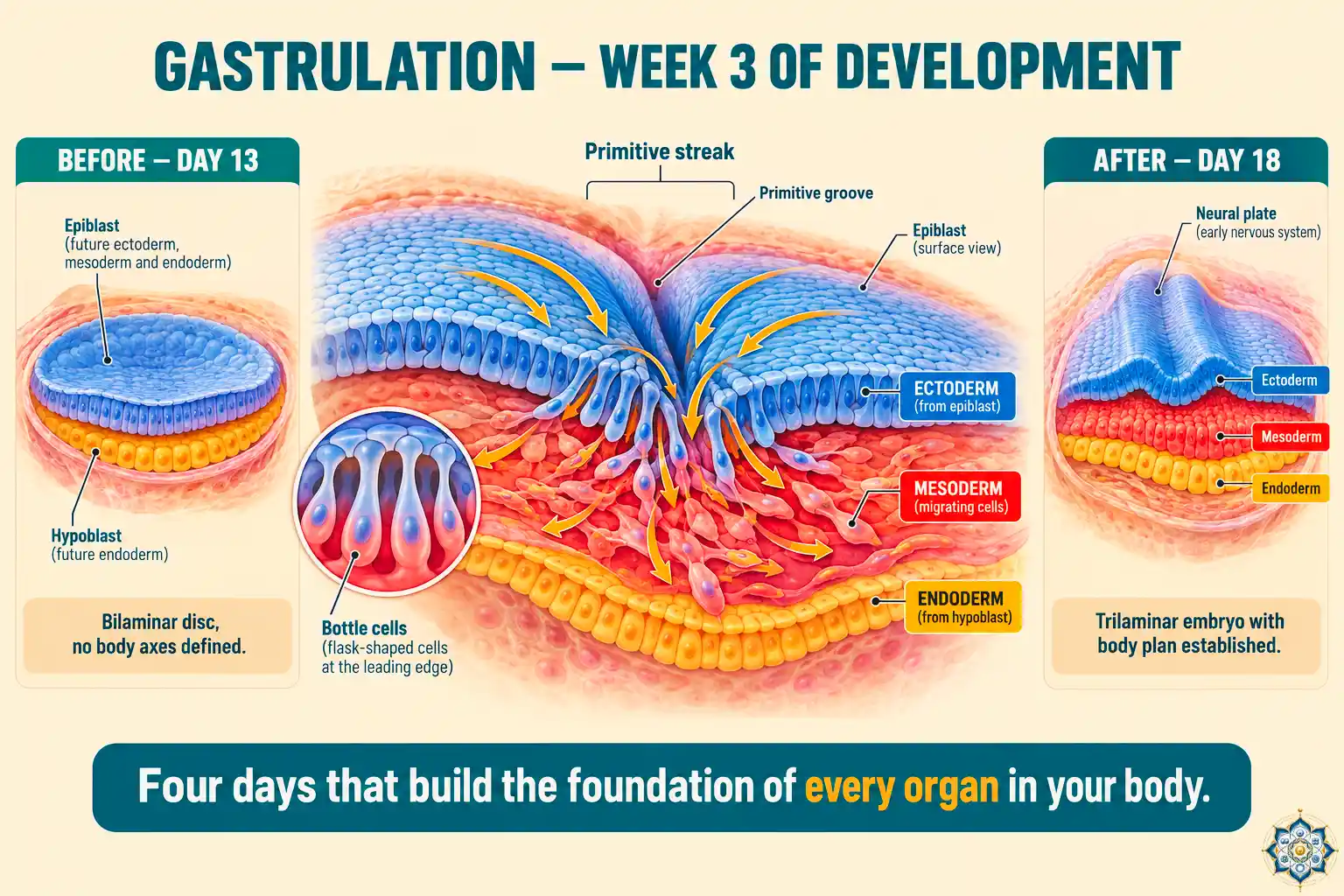
By the end of the second week after fertilization, the embryo has implanted in the uterine wall and has formed what’s called the bilaminar embryonic disc — literally, a two-layer plate.
The two layers are:
- Epiblast — the upper layer, made of tall columnar cells. This layer will give rise to the embryo proper (the actual body of the future person).
- Hypoblast — the lower layer, made of smaller cuboidal cells. This layer mostly contributes to extraembryonic structures like the yolk sac.
At this point, the embryo has no head, no tail, no front, no back, no left, no right. It’s a tiny circular plate about 0.2 mm across. Every cell in the epiblast is still essentially the same — undifferentiated, waiting for instructions.
Then, around day 14 after fertilization, something starts to form on the epiblast surface. A faint linear ridge appears. This is the primitive streak, and it’s the start of gastrulation.
The primitive streak: the embryo’s first axis
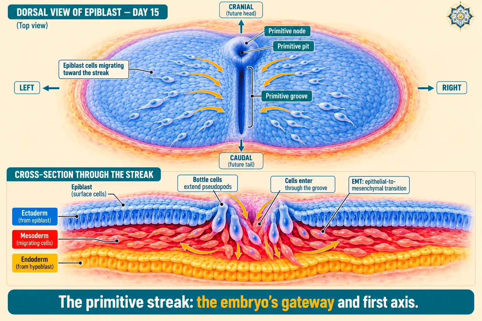
The primitive streak is the single most important landmark in gastrulation. Its appearance does several things simultaneously:
- It defines the body axis. The streak lies along what will become the midline from head to tail. Where the streak is, the back is; the opposite side is the belly.
- It creates bilateral symmetry. Everything that happens from now on is organized around this midline — your left arm mirrors your right arm because the streak set the mirror plane.
- It defines the head end. At one end of the streak sits the primitive node, a small mound of cells with a shallow depression called the primitive pit. This end becomes the head.
- It’s the gateway. Cells from the epiblast migrate toward the streak and ingress through it — moving inward from the surface to form the layers beneath.
How cells enter: bottle cells and pseudopods
At the primitive streak, the normally orderly sheet of epiblast cells does something unusual. Individual cells begin to change shape — their tops narrow while their bottoms stay wide. These are called bottle cells because of their flask-like appearance. They extend pseudopods (foot-like projections) downward, lose their connections to their neighbors, and crawl through the groove into the space below.
This single cellular trick is one of the most important in developmental biology. It’s a process called epithelial-to-mesenchymal transition (EMT) — a tightly connected sheet cell converts into a free-moving migratory cell. (You’ll meet EMT again in a moment — it’s one of the most reused tricks in biology.)
How the three germ layers form
Cells that enter through the primitive streak go to different places depending on when and where they ingress. The sequence is strict:
Wave 1: Endoderm
The first cells to migrate through the streak push downward and displace the hypoblast, forming the deepest layer. This becomes the endoderm (meaning “inner skin”). The endoderm will later form the lining of the digestive tract, the lungs, the liver, the pancreas, and parts of the bladder.
Wave 2: Mesoderm
The next wave of cells spreads out between the epiblast above and the newly-formed endoderm below. This middle layer becomes the mesoderm (“middle skin”). It’s the most diverse of the three layers and will give rise to muscles, bones, blood, kidneys, heart, blood vessels, and much more.
Wave 3: Ectoderm (what remains)
The cells that stay behind on the dorsal surface become the ectoderm (“outer skin”). This layer will form the entire nervous system (brain, spinal cord, nerves) and the outer skin layer (epidermis), including hair, nails, and tooth enamel.
By about day 17-18, the embryo has gone from a two-layer disc to a trilaminar (three-layer) embryo. Every one of the roughly 200 cell types in your adult body traces its ancestry back to one of these three layers.
What each germ layer becomes
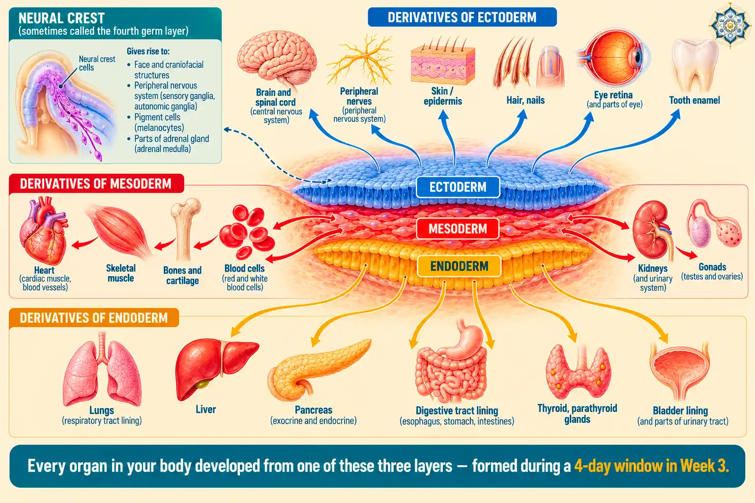
| Germ Layer | Major Derivatives |
|---|---|
| Ectoderm (outer) | Brain, spinal cord, peripheral nerves, retina, inner ear, epidermis, hair, nails, tooth enamel, mammary glands, pituitary gland (anterior) |
| Mesoderm (middle) | Skeletal muscle, smooth muscle, cardiac muscle, bones (except facial), cartilage, blood cells, blood vessels, lymphatic vessels, heart, kidneys, gonads, dermis of skin, spleen, adrenal cortex |
| Endoderm (inner) | Lining of digestive tract, lungs, liver, pancreas, thyroid, parathyroid, thymus, bladder lining, urethra lining, auditory tube |
The neural crest: the “fourth germ layer”
A special population of ectoderm cells at the border of the neural plate forms what some developmental biologists call the fourth germ layer: the neural crest. These cells migrate extensively throughout the embryo and give rise to a remarkable range of tissues — including most of the face, the peripheral nervous system, pigment cells (melanocytes), smooth muscle of major arteries, and parts of the adrenal gland. Vertebrates evolved the neural crest, and it’s largely responsible for the complexity of the vertebrate head.
The Nobel-prize-winning experiment that cracked gastrulation open
In 1924, two scientists in Freiburg, Germany performed an experiment that would reshape developmental biology forever.
Hans Spemann was a well-established embryologist. Hilde Mangold was his graduate student — a 24-year-old woman working on her PhD thesis. Together, they asked a simple question: if the dorsal lip of the blastopore (the region where gastrulation begins in a newt embryo) is transplanted to another embryo, what happens?
The experiment
Mangold took the dorsal lip tissue from a lightly-pigmented newt embryo and transplanted it into the belly region of a darkly-pigmented embryo. The pigment difference let her tell donor cells from host cells. She used extraordinarily primitive tools: glass needles made in the lab, and a loop of hair from Spemann’s child, sealed in a glass tube with wax, to manipulate the tissue.
Of over 250 embryos she operated on, only 6 survived long enough to show results. Those 6 changed biology.
The result
The surviving embryos developed two complete body axes. The host embryo had its normal head, body, and tail. But at the site of the transplant, a second complete embryo had formed — including a secondary neural tube, somites, and a brain — all built from host cells, not from the transplanted tissue.
The transplanted dorsal lip hadn’t just added tissue. It had instructed the surrounding host cells to become a new embryo. Spemann and Mangold had discovered what they called the organizer — a region of the embryo that tells all its neighbors what to become.
The tragedy and the Nobel Prize
Hilde Mangold died in 1924, the same year her thesis was published, at age 26, from burns suffered when a gasoline heater exploded in her kitchen. She never saw her discovery recognized.
In 1935, Hans Spemann was awarded the Nobel Prize in Physiology or Medicine for the discovery of the organizer effect. Nobel Prizes cannot be awarded posthumously, so Mangold’s name was not on the prize — but the Nobel Committee cited her thesis in its announcement. The organizer region is now universally called the Spemann-Mangold organizer in her honor.
The molecular organizer: what the signals actually are
For 60 years after Spemann and Mangold’s discovery, biologists knew the organizer existed but didn’t know how it worked. What were the signals? What were the molecules?
Starting in the 1990s, molecular biology finally cracked it. The organizer, it turns out, secretes a cocktail of signaling molecules — and more importantly, it secretes antagonists of other signals.
The key players
- Nodal — a TGF-β family signal essential for mesoderm formation and left-right asymmetry. Nodal gradients tell cells how far from the midline they are.
- BMP (Bone Morphogenetic Protein) — a signal that pushes cells toward ventral (belly) fates. High BMP = skin or blood. Low BMP = neural tissue.
- BMP antagonists: Chordin, Noggin, Follistatin — proteins secreted by the organizer that block BMP. Where these antagonists dominate, neural tissue forms. The organizer essentially creates a BMP-free zone, and that zone becomes the nervous system.
- Wnt signaling — establishes the anterior-posterior axis. High Wnt in the back, low Wnt in the front.
- FGF (Fibroblast Growth Factor) — drives the cell movements of gastrulation itself.
The organizer isn’t producing one magical “become an embryo” signal. It’s creating a gradient landscape — different signals at different concentrations in different regions. Each cell reads its position in the landscape and decides what to become based on that position.
This is one of the deepest ideas in developmental biology: cells don’t know what they are. They know where they are. Fate follows position.
The EMT trick that cancer later borrows
Remember the “bottle cells” that enter the primitive streak — cells that lose their connections to their neighbors, extend pseudopods, and crawl into the space below? That’s EMT (Epithelial-to-Mesenchymal Transition), and it’s one of the most important cellular tricks in biology.
EMT is used:
- In gastrulation — to form the mesoderm layer
- In neural crest migration — to disperse cells across the embryo
- In wound healing — to let epidermal cells crawl across a wound
- In cancer metastasis — to let tumor cells escape and spread
The same transcription factors (SNAIL, SLUG, TWIST, ZEB1) that drive EMT during gastrulation are the ones hijacked by cancer cells to metastasize. This is one of the most direct examples of how cancer is development gone wrong — tumors reactivating gastrulation’s cellular program in adult tissue.
Why gastrulation ends the twinning window
Before gastrulation, if an embryo splits into two pieces, each piece can develop into a complete individual. This is how identical twins form. If the split happens before day 8, the twins get separate placentas. If it happens between days 8-13, they share a placenta. If it happens between days 13-15, they share an amniotic sac.
But if a split happens after gastrulation begins (around day 15+), the resulting embryos can’t develop normally. The body axes have already been established. Splitting after that point produces conjoined twins — two bodies sharing some tissues because the gastrulation program only ran once.
Gastrulation effectively closes the window of embryonic flexibility. Before it, you could become two people. After it, you are definitively one — and whatever shape your gastrulation established, you will spend your life inhabiting.
Gastrulation across species: same event, different styles
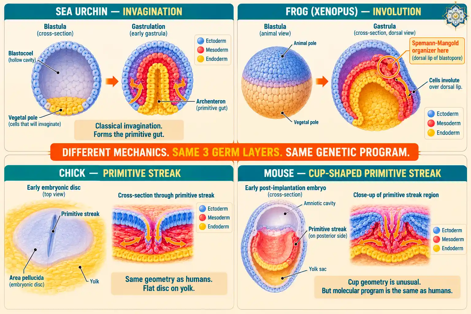
Every animal more complex than a sponge gastrulates. The specific mechanics vary dramatically, but the outcome is the same — a three-layer embryo with a body plan.
- Sea urchin — the classic model. Cells invaginate (push inward) at one pole to form the archenteron, the primitive gut tube. First-year biology textbooks still use this as the clearest example of gastrulation.
- Frog (Xenopus) — the organism Spemann and Mangold studied. Cells involute (roll inward) over the dorsal lip of the blastopore. The ‘yolk-heavy’ side moves differently than the lighter side.
- Chick — like humans, forms a primitive streak on a flat disc of cells sitting on top of yolk. Chicks are a key model for understanding mammalian gastrulation because the geometry is similar.
- Mouse — the embryo cups into a bowl shape before the primitive streak forms, which makes it look very different, but the underlying program is the same as in humans.
- Zebrafish — a modern favorite because the embryo is transparent, so cell movements can be watched live under a microscope.
The deep conservation across species is one of the central insights of evolutionary developmental biology. Different animals look wildly different, but the fundamental program of gastrulation — three germ layers, specified by shared toolkit genes — is nearly universal across the animal kingdom.
When gastrulation goes wrong: birth defects
Because gastrulation establishes the body plan, errors in it can produce some of the most striking congenital conditions:
- Sacrococcygeal teratoma — if the primitive streak doesn’t fully regress at the end of gastrulation, remnant pluripotent cells can form a tumor at the base of the spine containing teeth, hair, and other tissue types. It’s one of the most common tumors in newborns.
- Caudal regression syndrome — incomplete development of lower body structures caused by disrupted caudal gastrulation.
- Situs inversus — organs mirror-reversed because the left-right asymmetry signals around the primitive node failed. Usually harmless if all organs are mirrored (full situs inversus); dangerous if only some are (heterotaxy).
- Spina bifida (open neural tube defects) — technically a neurulation defect, but neurulation depends on signals set up during gastrulation. Folate deficiency during Week 3-4 of pregnancy is the strongest known risk factor.
This is why obstetricians recommend folate supplementation before conception. By the time most women know they’re pregnant (around 4-6 weeks after their last period), gastrulation is often already complete.
Why Wolpert said it was the most important time in your life
Lewis Wolpert’s famous quote — “It is not birth, marriage, or death, but gastrulation which is truly the most important time in your life” — isn’t hyperbole. It’s a precise statement about causality in biology.
Gastrulation is the moment when:
- Your body plan is set — head, tail, back, belly, left, right, all specified
- Every one of your future organs gets its origin layer
- The twinning window closes — you become definitively one person
- The cellular programs that will shape every tissue get activated
- Your bilateral symmetry gets established, including the subtle left-right asymmetries that put your heart on the left
Birth is the moment you’re separated from your mother. Marriage is a social event. Death is the end. But gastrulation is the moment you became you — the moment biology stopped treating you as a flexible cluster of cells and started building the specific body you’d inhabit for the rest of your existence.
And it happened in four days, when you were smaller than a grain of rice, before anyone — including your mother — knew you existed.
Key Takeaways
- Gastrulation is the embryonic event that transforms a two-layer disc of cells into a three-layer embryo with a defined body plan
- In humans, it occurs during Week 3 after fertilization (days 14-21), when most women don’t yet know they’re pregnant
- The primitive streak forms first, establishing the body axes and bilateral symmetry
- Cells migrate through the streak in three waves to form endoderm (first), mesoderm (second), and ectoderm (last)
- Every organ in your body derives from one of these three germ layers
- Hilde Mangold and Hans Spemann’s 1924 organizer experiment proved cells can instruct neighbors what to become — a discovery that won the 1935 Nobel Prize
- The molecular organizer works through signals (Nodal, BMP, Wnt, FGF) and antagonists (Chordin, Noggin, Follistatin) that create a gradient landscape cells read for position
- The EMT program used in gastrulation is reused by cancer cells to metastasize
- Gastrulation closes the twinning window — once it begins, splits produce conjoined twins rather than identical twins
Frequently Asked Questions
What is gastrulation in simple terms?
Gastrulation is the process by which a two-layer embryonic disc transforms into a three-layer embryo with a body plan. The three layers — ectoderm, mesoderm, and endoderm — are the sources of every tissue and organ in the adult body. In humans, gastrulation occurs during Week 3 of embryonic development.
When does gastrulation occur in humans?
Gastrulation begins around day 14-15 after fertilization and is largely complete by day 17-18. The primitive streak continues producing mesoderm until the end of Week 4. In pregnancy dating (counted from the last menstrual period), this corresponds roughly to weeks 4-5 — a time when most women don’t yet know they’re pregnant.
What are the three germ layers?
The three germ layers formed during gastrulation are ectoderm, mesoderm, and endoderm. Ectoderm forms the nervous system and skin. Mesoderm forms muscles, bones, blood, and most organs. Endoderm forms the lining of the digestive tract and respiratory system, plus the liver and pancreas. Every one of your ~200 cell types traces back to one of these three layers.
What is the primitive streak?
The primitive streak is a linear ridge that forms on the surface of the epiblast at the start of gastrulation. It establishes the embryo’s head-tail axis and bilateral symmetry. Cells from the surrounding epiblast migrate toward the streak, ingress through it, and form the three germ layers beneath. The primitive node at one end of the streak marks the future head region.
Who discovered the organizer region of the embryo?
Hilde Mangold and Hans Spemann discovered the organizer region in 1924 at the University of Freiburg. Mangold, working on her PhD thesis, transplanted the dorsal lip of the blastopore from a lightly-pigmented newt embryo into a darkly-pigmented one and found that a second complete embryonic axis formed from host cells. Spemann received the 1935 Nobel Prize for this work; Mangold died in 1924 at age 26 and was ineligible for the posthumous prize.
Why can’t identical twins form after gastrulation starts?
Before gastrulation, embryonic cells remain flexible enough that if the embryo splits, each half can regenerate a complete body plan. Once gastrulation begins and the body axes are established, splitting the embryo produces conjoined twins rather than identical twins — because the single body plan was already partially built. This is why identical twinning is limited to roughly days 1-14 after fertilization.
Follow HowLifeBuilds
@howlifebuilds