michael-levin-bioelectricity-revolution
Michael Levin bioelectricity research — the study of how electrical voltage patterns across cells direct body plan formation and regeneration — is one of the most provocative directions in modern developmental biology. It began with a single experiment that should have been impossible.
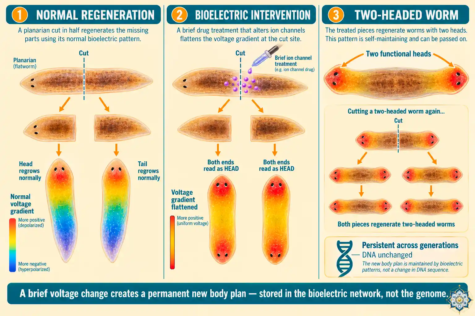
In 2007, Levin’s team at Tufts University took a flatworm — a tiny creature called a planarian, famous for its ability to regenerate — and cut it in half. Normally, the head half regrows a tail, and the tail half regrows a head. That’s the end of the story.
But Levin’s team didn’t let the biology play out normally. They briefly altered the bioelectric voltage of cells at the cut site — not with surgery, not with gene editing, just a quick chemical nudge to the cells’ membrane potential. The result was a worm with two heads. One at each end. Mirror symmetric. Fully functional.
Then they did something stranger. They cut these two-headed worms in half again. Both halves regrew into new two-headed worms. The altered body plan had been stored somewhere, and it kept getting read out through each new round of regeneration — even though the worms’ DNA was completely unchanged.
This single experiment hints at something that has rewritten what developmental biologists thought they knew about how bodies are built: DNA is not the only instruction set. There’s another layer — an electrical one — that tells cells where to go, what to become, and what shape the whole organism should take.
That electrical layer is what Michael Levin has spent two decades mapping. He calls it the bioelectric code, and if he’s right, it may be as fundamental to life as the genetic code itself.
The forgotten history of body electricity
The idea that life runs on electricity isn’t new. In the 1780s, Luigi Galvani made frog legs twitch by touching them with metal — the first demonstration that electrical signals exist in living tissue. Mary Shelley’s Frankenstein (1818) drew directly on this emerging science.
In the mid-20th century, Yale anatomist Harold Saxton Burr proposed that living things carry electrical “fields of life” that shape their development. Robert O. Becker, an orthopedic surgeon, spent decades documenting the electrical patterns at wound sites and limb stumps in salamanders. His 1985 book The Body Electric argued that bioelectric signals control regeneration — and that humans had quietly lost the ability to listen to them.
Then the field went quiet. The DNA revolution had arrived. Molecular biology took center stage. Genes became the universal explanation for everything from eye color to behavior. Bioelectricity was pushed to the margins — treated as either a side effect of life, or as a signal only relevant to nerves and muscles.
Levin’s work has resurrected it as something far bigger.
What is the bioelectric code?
At the heart of Michael Levin bioelectricity research is a simple observation: every cell in your body is a tiny battery.
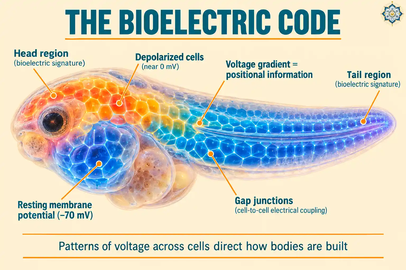
The membrane of each cell separates charged ions — mostly sodium, potassium, calcium, and chloride — between the inside and the outside. The imbalance creates a voltage across the membrane called the resting membrane potential, typically ranging from –90 mV (strongly negative inside) to near 0 mV (depolarized) depending on cell type.
Neurons use fast, dramatic voltage spikes to send signals. That’s familiar. What Levin’s work has shown is that every cell — not just nerve cells — carries a voltage, and these voltages talk to each other through gap junctions (tiny channels that physically connect neighboring cells).
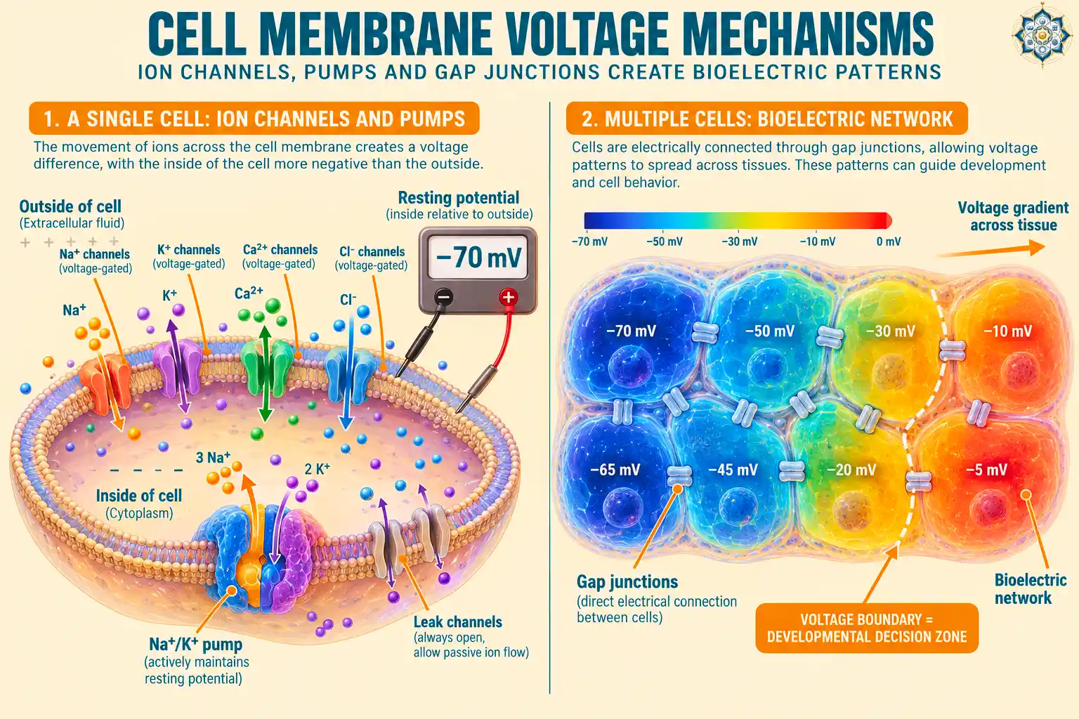
Together, millions of connected cells form what Levin calls bioelectric networks. The pattern of voltages across these networks — the places where voltage is high, low, or changing — forms a kind of electrical map. And that map appears to tell cells things like:
- Where the head goes and where the tail goes
- Which side is left and which is right
- Where to grow an eye, a limb, or a heart
- When to stop growing
Cells don’t just read these patterns — they can rewrite them. The system is dynamic, and it operates at a higher level of abstraction than the genes acting underneath it.
Gradients, not absolute voltages
One of Levin’s key insights is that cells don’t respond to a single voltage value — they respond to differences in voltage between regions. A gradient, in other words. Many developmental decisions happen at the boundaries where one voltage domain meets another.
This is important because it means the system is self-correcting at a scale larger than any single cell. The whole network interprets the shape of the voltage landscape, not individual readings.
The two-headed planaria — a signal that outlasts the signal
The planarian experiment is the clearest demonstration of what the bioelectric code can do.
The protocol was simple: Levin’s team cut planarians, then briefly treated the regenerating cells with drugs that opened or closed specific ion channels, shifting the voltage pattern at the wound site. In worms where the voltage gradient was flattened — making both ends look electrically similar to a “head region” — the worms regenerated with heads at both ends.
The researchers didn’t change a single gene. They didn’t add any DNA. They just briefly altered the voltage pattern, and the body plan changed.
Then came the stranger part: when they cut the two-headed worms again, with no further treatment, the pieces regenerated into more two-headed worms. The altered pattern had become the new instruction set. As Levin put it: “If you recut them, they make more two-headed worms.”
Growing an eye where no eye belongs
If voltage patterns can change where a head goes, could they also create organs in places they don’t normally form?
In 2011, Levin’s team tested this on tadpoles. They identified the specific voltage pattern that normally sets up the eye field — the region of embryonic tissue that will become the eye. Then they recreated that voltage pattern in a completely different part of the embryo: the gut.
They did this by injecting mRNA that caused gut cells to express a single additional ion channel, shifting those cells’ voltage to match what the eye field would normally look like.
A functional eye grew on the tadpole’s gut.
Lens. Retina. Pigmented epithelium. All of it, from gut tissue that had no business becoming an eye. The gut cells recognized the voltage pattern as a “build an eye” instruction and executed it using whatever genetic machinery they had available.
This result changed how Levin thinks about cells. They aren’t locked into a single fate by their DNA. They carry latent competencies — abilities to become many different things — that specific bioelectric signals can switch on. The DNA provides the hardware. The bioelectric code decides what software runs.
Six-legged frogs and the master channels
Levin’s team has also produced frogs with extra limbs by manipulating voltage gradients at the right locations during development. These aren’t deformed stumps — they’re fully functional legs with proper bone, muscle, and nerve organization.
What makes this possible is a striking simplicity in how bioelectric control is organized:
- In most cells, only one or two ion channels dominate the resting voltage. Change those few channels, and you shift the entire cell’s electrical identity.
- Just four ion channels appear to act as master controls for left-right organ placement across the whole body. Alter any one of them, and the embryo randomizes which side the heart, liver, and stomach end up on.
This hints at something extraordinary: large-scale body plan decisions may be governed by a small number of key electrical switches, not by thousands of individually programmed cells.
How you read and write bioelectric signals
Levin’s lab developed the toolkit that made this field possible. The approach has two sides:
Reading voltages
Voltage-sensitive fluorescent dyes change their brightness or color depending on membrane potential. By soaking embryos or tissue in these dyes and imaging them with a microscope, researchers can see the voltage pattern across every cell — producing a glowing map of the bioelectric landscape.
Writing voltages
To change the pattern, Levin’s team uses several tools:
- Ion channel drugs — small molecules that open or close specific channels
- mRNA injection — forcing specific cells to express extra ion channels, shifting their voltage
- Light-sensitive channels (optogenetics) — allowing precise control of voltage with a beam of light
- Ionophores — molecules that carry specific ions across membranes
These tools let the researchers both observe what voltage pattern a tissue is holding, and rewrite it to see what happens — a hardware debugger for development.
Xenobots: the first living robots
In 2020, Levin and roboticist Josh Bongard at the University of Vermont published something that didn’t quite fit any existing category.
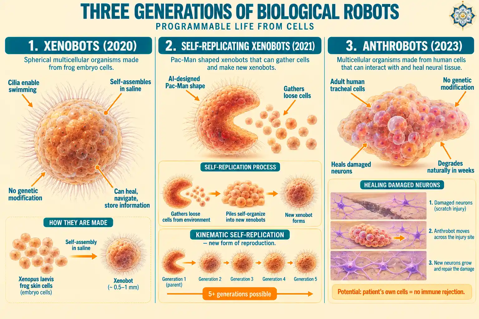
They took skin stem cells from African clawed frog (Xenopus laevis) embryos — just the cells, no embryo, no genes added — and placed them in a saline bath. The cells did something remarkable on their own: they clustered into spheres, grew cilia (tiny hair-like structures), and started swimming.
These weren’t frogs. They weren’t embryos. They were a new kind of multicellular organism that had never existed in nature. The researchers called them xenobots.
The original xenobots could:
- Swim through liquid using coordinated cilia
- Navigate narrow passageways
- Push particles into piles
- Heal themselves after being cut
- Store information from their experiences
The 2021 breakthrough: self-replication
In November 2021, the team announced something even stranger. Using an evolutionary algorithm running on a supercomputer, they simulated billions of possible xenobot shapes to find one that could reproduce. The winning design looked like Pac-Man — a sphere with a wedge cut out.
When these Pac-Man xenobots were placed in a dish with loose frog stem cells, they used their open mouths to gather the loose cells into piles. Those piles then self-organized into new spherical xenobots. A single Pac-Man xenobot could trigger at least five generations of offspring.
This is a form of reproduction — called kinematic self-replication — that had never been observed in any animal or plant. Molecules replicate this way. Cells don’t. Until now.
Anthrobots (2023): human cells can do it too
In late 2023, Levin’s lab announced anthrobots — biological robots built from adult human tracheal (windpipe) cells, with no genetic modification.
Anthrobots self-assemble from single cells into multicellular structures ranging from the width of a human hair to the tip of a sharpened pencil. They move across surfaces using cilia. And in the most striking experiment, researchers scratched a layer of human neurons to create damage, then released anthrobots onto the scratched surface. The anthrobots moved across the gap — and encouraged new neuron growth to fill the damaged region.
How they do this is still being investigated. But the implication is enormous: because anthrobots can be made from a patient’s own cells, they would likely avoid immune rejection, and they naturally break down within weeks.
What this could mean for medicine
If Michael Levin bioelectricity research proves out at scale, several areas of medicine could change radically — not someday, but within the next decade or two.
Limb and organ regeneration
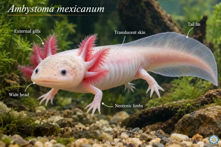
Humans retain most of the regeneration genes that axolotls use to regrow entire limbs — we just don’t activate them. Levin’s work suggests we might be able to switch them on by recreating the right bioelectric environment at a wound site.
His team, in collaboration with David Kaplan at Tufts, developed a device called the BioDome — a watertight silicone and silk wearable that creates a controlled electrochemical environment over a wound. In tadpoles, a one-hour BioDome treatment at the amputation site produced a fully regrown tail in about eight days. A human amputee might one day wear a similar device for a few hours to kickstart limb regrowth.
Cancer
Levin suspects that cancer may partly be a bioelectric failure — cells become abnormally depolarized, lose their place in the voltage network, and start behaving as individuals rather than as part of a tissue. In frog experiments, his team used light-activated ion channels to reverse tumors by restoring normal bioelectric signaling, without chemotherapy.
If this translates to humans, the goal wouldn’t be killing cancer cells. It would be convincing them to rejoin the tissue. This fits a broader reframing in cancer biology: cancer is development gone wrong, cells reverting to an inappropriately embryonic state. Bioelectric correction may be one of the fastest routes to reversing that reversion.
Birth defect correction
Levin’s team has demonstrated that bioelectric interventions in developing frog embryos can reverse malformations — including a forebrain defect that mirrors conditions linked to parental alcohol exposure in humans. If prenatal screening could detect bioelectric abnormalities early, drugs that target ion channels (many of which already exist for heart and neurological conditions) might correct developmental errors before birth.
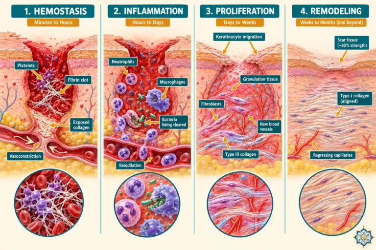
Scar-free wound healing
The same voltage signals that direct regeneration also influence how human wounds heal and whether they scar. Shifting the bioelectric response at a wound site could, in principle, nudge human healing toward regeneration rather than fibrosis.
Where the skeptics push back
Not everyone in developmental biology is convinced that bioelectricity is as central as Levin’s work suggests.
Andre Levchenko (Yale) has argued that the field needs stronger mechanistic understanding before accepting bioelectricity as a primary driver of development — rather than a downstream readout of genetic signaling. Cliff Tabin (Harvard) has questioned whether ion channels truly control developmental decisions or whether they’re correlated with decisions driven by other molecular pathways.
These are legitimate concerns. Correlation isn’t causation, and the field is still working out exactly how the voltage pattern connects downward to gene expression and upward to tissue-level behavior.
Levin’s own position is that bioelectricity isn’t a replacement for genetics — it’s a complementary layer. DNA is the hardware. Bioelectric networks are something closer to the operating system that decides how the hardware runs. Both are needed. Neither is sufficient alone. The genetic layer itself is largely the evo-devo toolkit — a small set of ancient genes like Hox and Pax6 that every animal uses — and bioelectric signals appear to be one of the ways cells decide which toolkit genes to activate where.
Why this changes how we think about life
For most of the last 70 years, biology has operated on an implicit assumption: DNA tells cells what to do. Development, regeneration, aging, disease — all were seen primarily as consequences of which genes get expressed, when, and in which cells.
Michael Levin bioelectricity work doesn’t deny any of that. But it adds a layer on top: cells also talk to each other through electrical signals, those signals form patterns that span whole tissues, and those patterns carry instructions about shape, position, and identity that the genes alone don’t contain.
If this is right, then:
- Regeneration isn’t a matter of “turning on” lost genes — it’s a matter of restoring the right bioelectric pattern that tells the existing genes what to build
- Cancer isn’t just a genetic disease — it’s partly a breakdown of tissue-level electrical coordination
- Birth defects can sometimes be reversed by changing the voltage landscape, not just the genetic one
- Cells carry far more organizational potential than we assumed — the frog cells that became xenobots had always been able to do that, we just never gave them the chance
Levin’s framing is that life is a form of scalable intelligence. Individual cells have limited information, but they aggregate into larger decision-making units through bioelectric networks. Those networks solve problems — figuring out how to build a correctly-shaped body, how to heal an injury, how to maintain function over time — in ways that look increasingly like cognition.
“We are communicating to the cellular collective, and we talk about very abstract, high-level things.” — Michael Levin
Key Takeaways
- Every cell carries a voltage across its membrane; those voltages form networks that span whole tissues via gap junctions
- Patterns of voltage — the “bioelectric code” — direct where organs form, how body plans are built, and how regeneration proceeds
- Levin’s team produced two-headed planaria, extra limbs in frogs, and eyes in gut tissue by manipulating voltage patterns alone — without genetic changes
- Xenobots (2020, 2021) and anthrobots (2023) show that cells carry latent organizational competencies far beyond their default role
- Clinical applications in development include limb regeneration (BioDome device), tumor reversal, birth defect correction, and scar-free wound healing
- Bioelectricity complements genetics rather than replacing it — DNA is the hardware, bioelectric networks are closer to the operating system
Frequently Asked Questions
Who is Michael Levin?
Michael Levin is the Vannevar Bush Professor of Biology at Tufts University and director of the Allen Discovery Center at Tufts since 2016. He’s also an Associate Faculty member at Harvard’s Wyss Institute. His lab studies bioelectric signaling in development, regeneration, and cancer, and has pioneered the creation of biological robots called xenobots and anthrobots.
What is the bioelectric code?
The bioelectric code is the term Levin uses for the patterns of electrical voltage that cells maintain across their membranes, which collectively form a kind of map that directs body plan formation and regeneration. Unlike nerve signals (which are fast and transient), these are stable voltage differences that persist across tissues and encode instructions about shape, position, and cell identity.
How are the two-headed planarians produced?
Levin’s team briefly treats regenerating planarian flatworms with drugs that modify specific ion channels at the cut site. This flattens the normal voltage gradient between the head and tail regions, so both ends of the regenerating fragment “read” as head territory. The resulting two-headed worms produce more two-headed worms when cut again, even without further treatment, suggesting the altered pattern becomes stored in the bioelectric network itself.
What are xenobots?
Xenobots are tiny multicellular organisms built from skin stem cells of African clawed frog embryos. Placed in saline, these cells self-assemble into spheres covered with cilia that can swim, navigate, heal themselves, and in a 2021 Pac-Man shaped version — self-replicate by gathering loose cells into new xenobots. They’re the first entities that are neither traditional organisms nor machines.
What are anthrobots and how are they different?
Anthrobots are biological robots built from adult human tracheal cells, announced by Levin’s lab in late 2023. Unlike xenobots (made from embryonic frog cells), anthrobots use adult human cells with no genetic modification. In lab experiments, they moved across scratched layers of human neurons and encouraged new neuron growth, suggesting potential applications in healing damaged tissue. They break down naturally within weeks.
Could bioelectricity actually help humans regrow lost limbs?
Levin’s team, in collaboration with Tufts engineer David Kaplan, developed the BioDome — a wearable silicone and silk device that creates a controlled electrochemical environment over a wound. In frogs and tadpoles, a brief BioDome treatment triggered full regeneration of limbs and tails. Translating this to humans faces real obstacles (higher blood pressure, warm-blooded physiology, infection risk), but Levin believes clinical movement is possible within the next decade.
Follow HowLifeBuilds
@howlifebuilds