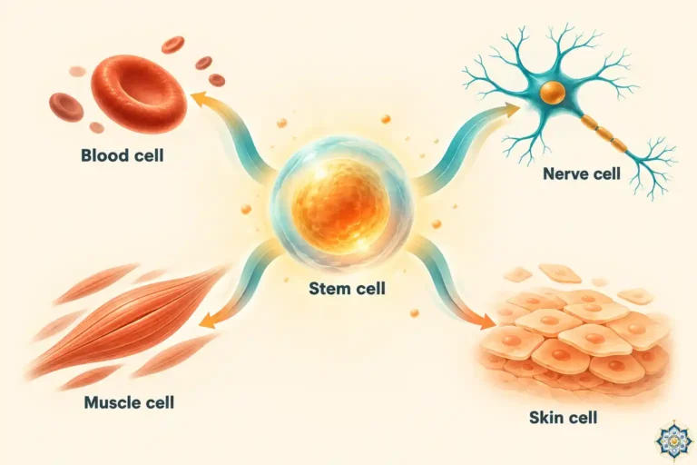
The First 24 Hours: What Happens After Fertilization
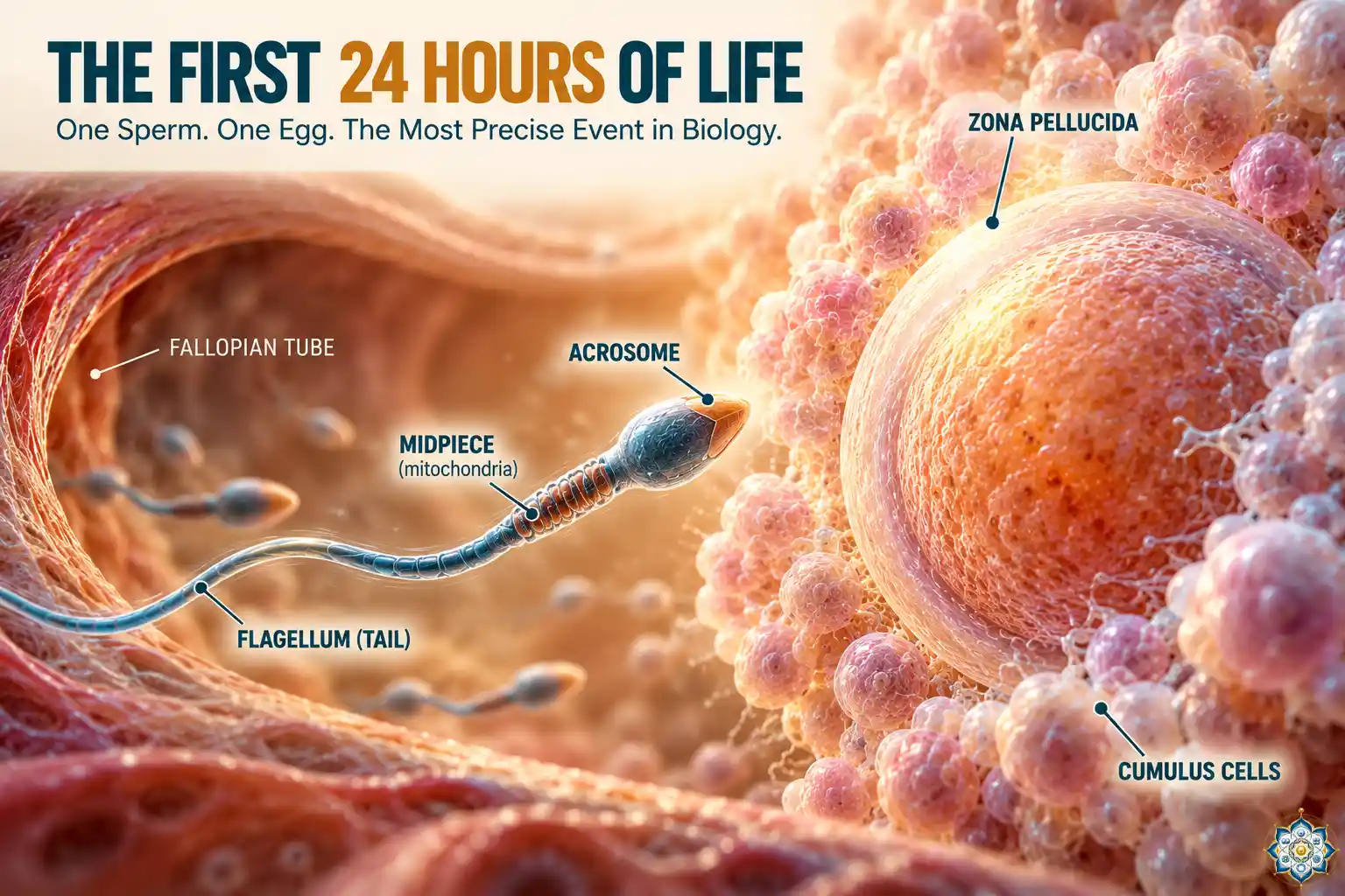
Within 24 hours of a single sperm penetrating the egg, the fertilized cell completes a staggering sequence of events: it blocks all other sperm, merges two half-genomes into one complete set of 46 chromosomes, decides the biological sex of the future person, reprograms its entire molecular machinery, and divides into the first two cells of a new human being. Every step is timed to the minute, and a failure at any stage ends the process.
What happens after fertilization? Think about the most precisely engineered machine you’ve ever seen. A Swiss watch. A spacecraft docking system. A Formula 1 engine. Now consider this: the first 24 hours of the human fertilization process make all of those look crude.
Coming from a background in mechanical engineering, I used to think of precision as something humans built. Then I started studying developmental biology, and I realized that the most astonishing engineering on Earth happens in a space smaller than the period at the end of this sentence.
In the next 16 minutes, I’ll walk you through exactly what happens after fertilization — minute by minute, molecule by molecule — from the instant a sperm touches the egg to the moment that single cell divides into two. This is where every human life starts — and what happens after fertilization is far stranger and more elegant than most people know.
Already pregnant and want to know exactly how far along you are? Use our free pregnancy due date calculator to find your EDD, current trimester, and conception date in seconds.
Key takeaways
- Of roughly 300 million sperm released, only about 200 reach the egg — a survival rate below 0.0001%.
- Sperm must spend 7–10 hours capacitating inside the female tract before they can fertilize an egg.
- The egg blocks additional sperm within seconds using a fast electrical block and a slower cortical reaction.
- The parental genomes don’t “fuse” — they remain in separate pronuclei for hours before combining on the first mitotic spindle.
- Biological sex is decided at the instant of fertilization by whether the sperm carries an X or Y chromosome.
- The first cell division happens at roughly 24–30 hours, creating a two-cell embryo.
- More than 50% of fertilized eggs fail to develop past the first week, most due to chromosomal errors.
- The 300 Million to 200 Problem — How Sperm Reach the Egg
- Capacitation — Why Sperm Aren’t Ready Yet
- First Contact — The Zona Pellucida and the Acrosome Reaction
- Sperm-Egg Fusion — The Moment of Fertilization
- Blocking Polyspermy — How the Egg Shuts the Door
- Two Pronuclei, One Cell — Combining Two Genomes
- X or Y — How Sex is Decided
- The Great Reprogramming — Resetting the Cellular Machinery
- The First Division — From One Cell to Two
- What Happens After Fertilization — Minute-by-Minute Timeline
- What IVF Teaches Us About Fertilization
- Frequently asked questions
1. The 300 Million to 200 Problem — How Sperm Reach the Egg
Let’s start with a number that puts everything in perspective. During ejaculation, roughly 200–300 million sperm are released. Of those, approximately 200 will reach the egg waiting in the fallopian tube. That’s a filtration rate of 99.9999%.
The journey from the cervix to the egg is about 15–18 centimeters — which doesn’t sound far until you realize that a sperm cell is only 50 micrometers long. Scaled up, that’s like a human swimming from London to New York.
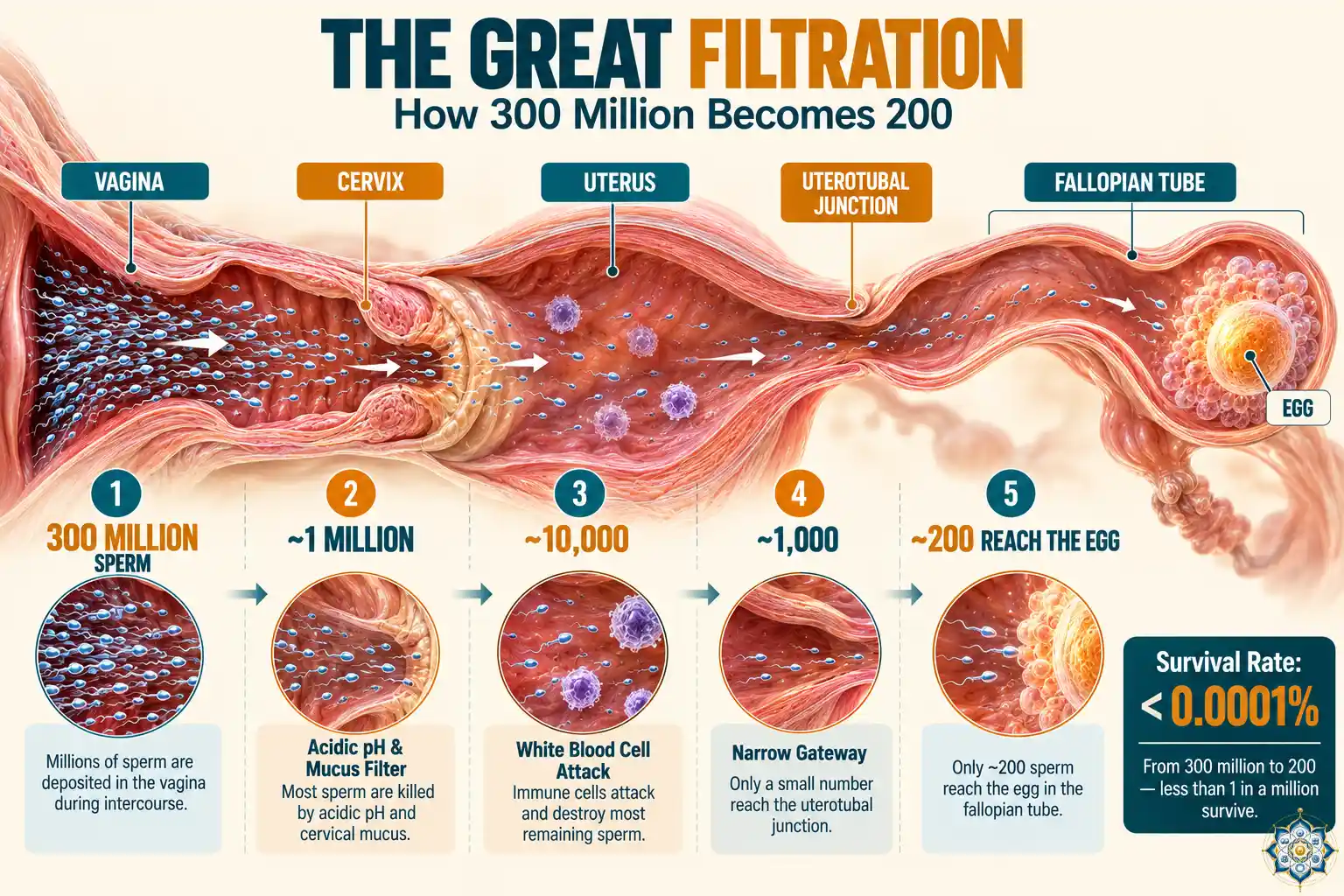
Here’s where the filtration happens:
| Stage | Location | Approx. sperm remaining | What eliminates them |
|---|---|---|---|
| Start | Vagina | 200–300 million | — |
| 1st barrier | Cervix | ~1 million | Acidic pH (~4.0), cervical mucus filtration |
| 2nd barrier | Uterus | ~10,000 | White blood cell attack (phagocytosis) |
| 3rd barrier | Uterotubal junction | ~1,000 | Narrow anatomical gateway |
| 4th barrier | Fallopian tube (wrong side) | ~500 | 50% swim into the wrong tube |
| Arrival | Egg (ampulla region) | ~200 | Final cumulus cell barrier |
This isn’t just wasteful — it’s quality control. The female reproductive tract functions as a sophisticated selection system. Sperm with abnormal morphology, weak motility, or damaged DNA are filtered out at each stage. By the time the survivors reach the egg, they represent the most motile, structurally sound sperm from the original population.
Why so many? The massive number of sperm isn’t about competition for the egg — it’s about overcoming the odds. Each barrier is a filter, and without the initial volume, not enough sperm would survive to guarantee that even one reaches the egg. Fertility problems often begin when the starting count drops below 15 million per milliliter.
2. Capacitation — Why Sperm Aren’t Ready Yet
Here’s something that surprises most people: freshly ejaculated sperm cannot fertilize an egg. They need to undergo a series of biochemical changes called capacitation, which takes 7–10 hours inside the female reproductive tract.
Capacitation involves three key changes:
Membrane destabilization. Cholesterol is removed from the sperm’s outer membrane, making it more fluid and fragile. This is necessary for the acrosome reaction later, but it also means capacitated sperm have a shorter lifespan — they’ve traded durability for function.
Calcium influx. Ion channels open, allowing calcium to flood into the sperm cell. This calcium surge is what drives the dramatic change in swimming pattern described below.
Hyperactivation. The sperm’s tail movement changes from a symmetrical, wave-like beat (good for straight-line swimming) to an asymmetrical, whip-like thrash. This hyperactivated motility generates the physical force needed to push through the egg’s outer layers. Under a microscope, the difference is dramatic — hyperactivated sperm look almost frantic compared to the smooth motion of uncapacitated ones.
This is why timing matters in reproduction. Sperm can survive in the reproductive tract for up to 5 days, but they only remain capacitated and capable of fertilization for about 12–24 hours. The egg, meanwhile, is only fertilizable for about 12–24 hours after ovulation. The window where everything overlaps is remarkably narrow.
3. First Contact — The Zona Pellucida and the Acrosome Reaction
The egg doesn’t sit naked in the fallopian tube. It’s surrounded by two layers of protection:
The cumulus cell layer — a cloud of sticky cells embedded in a hyaluronic acid matrix. This is the first obstacle a sperm must navigate. Enzymes on the sperm’s surface (and the physical force of hyperactivated swimming) help it push through.
The zona pellucida — a thick glycoprotein shell roughly 13 micrometers thick. This is the egg’s real gatekeeper. It contains four key glycoproteins (ZP1–ZP4), and one of them — ZP3 — is the receptor that recognizes sperm.
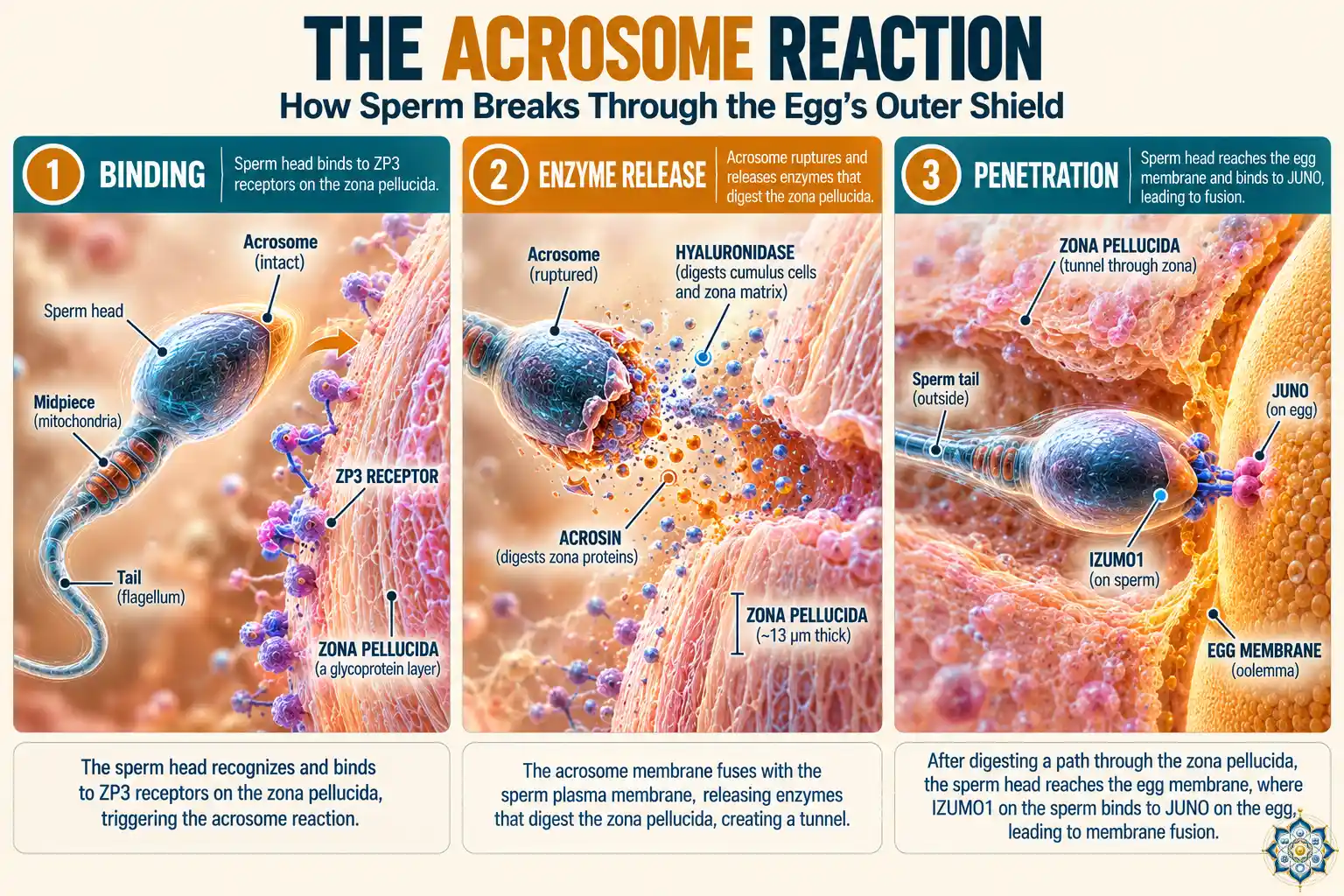
When a capacitated sperm binds to ZP3, it triggers the acrosome reaction — one of the most dramatic events in cell biology. The acrosome, a cap-like vesicle covering the front half of the sperm head, ruptures and releases a cocktail of digestive enzymes:
Hyaluronidase breaks down the hyaluronic acid in the cumulus layer. Acrosin (a protease) digests the glycoproteins of the zona pellucida. Together, these enzymes carve a narrow tunnel through the egg’s outer coat while the hyperactivated tail provides the driving force from behind.
The sperm doesn’t blast through — it drills. The process takes about 15–20 minutes, and the tunnel is barely wider than the sperm head itself.
Species specificity: ZP3 is one of the reasons cross-species fertilization almost never works. The ZP3 glycoprotein is species-specific — human sperm can only bind to human ZP3. It’s a molecular lock-and-key system that has evolved independently across mammalian lineages.
4. Sperm-Egg Fusion — The Moment of Fertilization
Once through the zona pellucida, the sperm reaches the egg’s plasma membrane. What happens next is not what most people picture.
The sperm doesn’t pierce the membrane like a needle. Instead, proteins on the sperm surface — primarily IZUMO1 — bind to a receptor on the egg membrane called JUNO. This receptor pair was only discovered in 2005 (IZUMO1) and 2014 (JUNO), and their names tell a story: IZUMO is named after a Japanese marriage shrine, and JUNO after the Roman goddess of marriage.
When IZUMO1 binds JUNO, the two cell membranes fuse. The sperm’s nucleus, mitochondria, and centriole are pulled into the egg’s cytoplasm. The sperm’s tail and most of its other structures are left behind or degraded.
And here’s a detail that matters: the sperm’s mitochondria — its energy-producing organelles — are specifically targeted for destruction inside the egg. They are tagged with ubiquitin (a “destroy this” molecular label) and broken down. This is why mitochondrial DNA is inherited exclusively from the mother. Every mitochondrion in your body traces back to your mother’s egg cell.
5. Blocking Polyspermy — How the Egg Shuts the Door
The moment the first sperm fuses with the egg membrane, a race begins — against every other sperm still approaching. If a second sperm enters, the resulting cell would have 69 chromosomes instead of 46 (a condition called triploidy), which is almost always fatal.
The egg has two defenses, and they operate on different timescales:
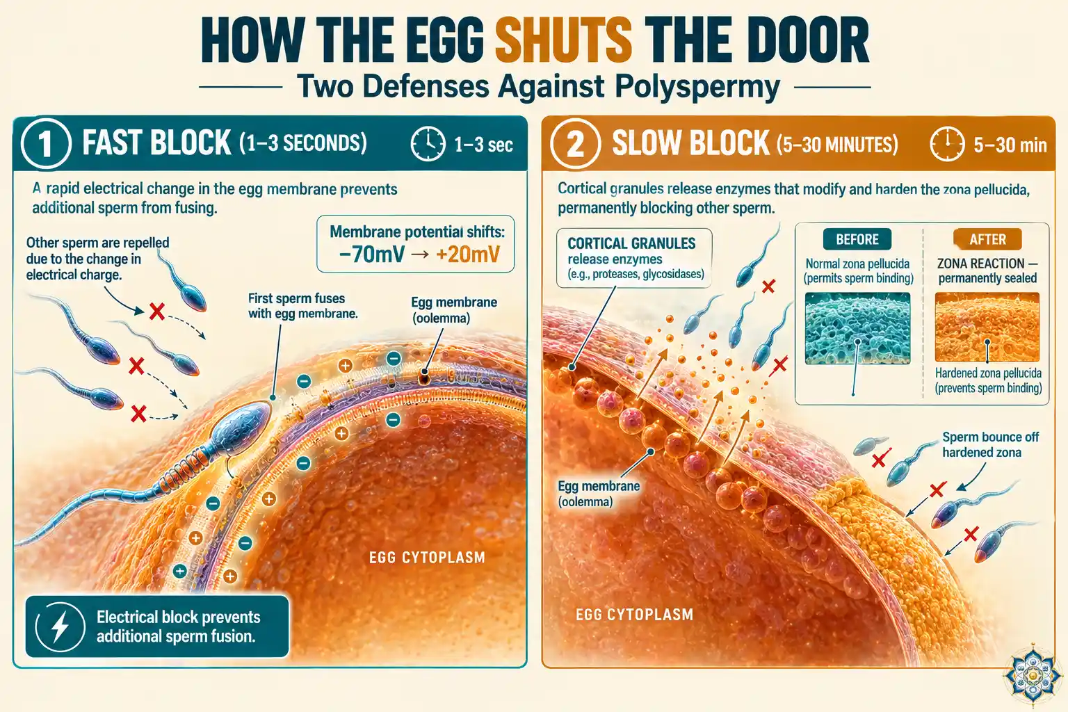
The fast block (1–3 seconds)
Within 1–3 seconds of sperm fusion, the egg’s membrane potential shifts from about –70 millivolts to +20 millivolts. This electrical change is too brief and too subtle to be visible, but it immediately prevents any additional sperm from fusing with the membrane. It’s the same principle as an electrical fence — except it operates at the scale of a single cell.
The slow block — the cortical reaction (minutes)
This is the permanent seal. Thousands of cortical granules — small vesicles just beneath the egg’s surface — fuse with the plasma membrane and dump their contents into the space between the membrane and the zona pellucida. These enzymes do two things:
They cleave ZP2, which destroys the sperm-binding sites. And they cross-link the zona’s proteins, physically hardening it into what’s called the zona reaction. The once-penetrable coat becomes a rigid shell. Any sperm still trying to drill through finds an impenetrable wall.
The JUNO receptor itself is also shed from the egg’s surface within about 40 minutes — so even if a sperm reached the membrane, there would be nothing to bind to.
6. Two Pronuclei, One Cell — Combining Two Genomes
Now comes the part that most biology classes get wrong. The sperm and egg nuclei don’t simply “merge.” What actually happens is more complex and far more interesting.
After the sperm enters, its tightly compacted DNA must be unpacked. In the sperm, DNA is wound around proteins called protamines (not histones, like in most cells), which compress it to about 1/6th the volume of a normal cell nucleus. The egg’s cytoplasm strips away these protamines and replaces them with histones, allowing the DNA to decondense and become readable again. This process takes several hours.
Meanwhile, the egg completes its own final cell division — meiosis II. This is a detail most people don’t know: at the time of fertilization, the egg hasn’t even finished dividing. The sperm’s entry triggers the completion of meiosis, producing the mature egg nucleus and a tiny discarded cell called the second polar body.
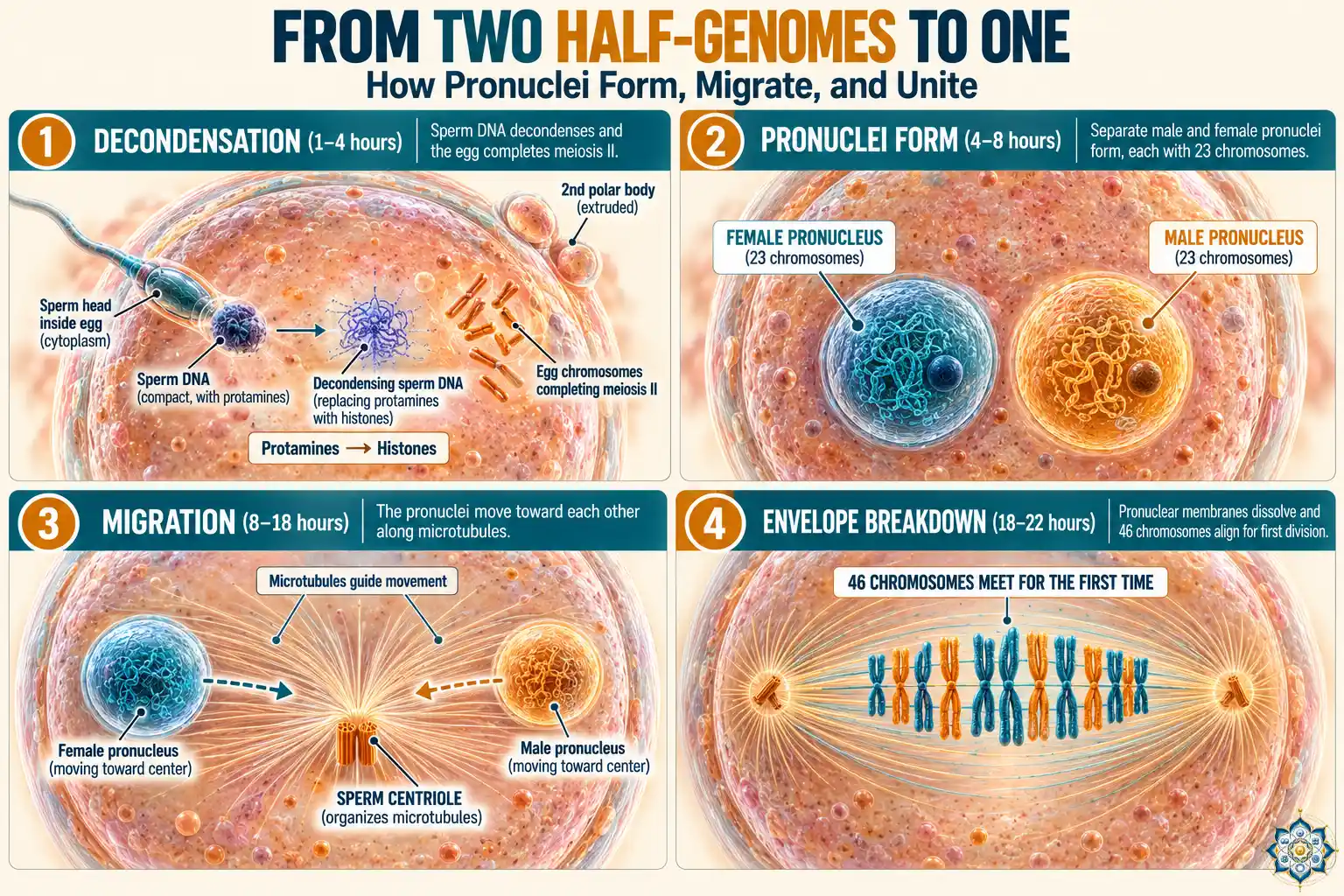
Once both DNA sets are decondensed and wrapped in fresh nuclear envelopes, they are called pronuclei:
The female pronucleus contains 23 chromosomes from the mother. The male pronucleus contains 23 chromosomes from the father. For several hours, these two pronuclei sit side by side in the same cell — visible under a microscope as two distinct circles. In IVF clinics, the observation of two pronuclei (called “2PN”) at 16–18 hours after insemination is the standard confirmation that fertilization has occurred normally.
The two pronuclei migrate toward the center of the cell, guided by microtubules organized by the sperm’s centriole (the egg’s own centrioles were lost during its development — this is one of the critical contributions the sperm makes beyond its DNA). Their nuclear envelopes break down. And for the first time, all 46 chromosomes line up together on the mitotic spindle.
This is the moment. Not a fusion of nuclei, but a choreographed demolition and reassembly — two separate genomes meeting on the same scaffold for the first time.
7. X or Y — How Sex is Decided
Every egg carries one X chromosome. Sperm carry either an X or a Y. The biology is simple. The consequences are not.
| Sperm type | Egg | Result | Outcome |
|---|---|---|---|
| X-bearing sperm | X | XX | Female |
| Y-bearing sperm | X | XY | Male |
This is decided at the instant the sperm enters the egg, and it cannot be changed afterward. No diet, no timing method, no position has been scientifically shown to reliably influence whether an X or Y sperm reaches the egg first. Despite decades of folk wisdom, the natural ratio remains approximately 105 males born per 100 females worldwide.
But the Y chromosome does more than just determine sex. It carries the SRY gene (Sex-determining Region Y), which won’t activate until about 6–7 weeks of embryonic development. When it does, it triggers a cascade that converts the default gonadal tissue into testes. Without SRY, the same tissue develops into ovaries. But all of that is weeks away. In the first 24 hours, the Y chromosome is just another piece of DNA being unwound and prepared for its first replication.
8. The Great Reprogramming — Resetting the Cellular Machinery
While the pronuclei are forming and migrating, something less visible but equally important is happening. The egg is reprogramming the sperm’s DNA — and its own.
Both the sperm and egg are highly specialized cells. The sperm is built for swimming and nothing else. The egg is loaded with molecular supplies but frozen in a suspended state. To produce an embryo capable of building every cell type in the human body, these specializations must be erased.
This involves epigenetic changes on a massive scale:
DNA demethylation. Methyl groups — chemical tags that silence genes — are stripped from the sperm’s DNA almost entirely within hours. The egg’s DNA undergoes demethylation too, but more gradually. This erasure is the reset button that converts two specialist cells into one totipotent cell — a cell capable of becoming anything.
Histone modification. After protamines are replaced with histones, these histones receive specific chemical modifications that open up the chromatin structure, making genes accessible for the embryo’s own developmental program.
Maternal mRNA activation. The egg contains stockpiles of messenger RNA molecules made during its growth phase. These maternal mRNAs are the embryo’s first instruction manual — they control everything until the embryo’s own genome switches on (which doesn’t happen until the 4–8 cell stage in humans, a process called embryonic genome activation). The genes that run this early program — pluripotency factors like OCT4, SOX2 and NANOG — are supposed to go silent after development completes. When they reactivate decades later in adult tissue, the consequence is often cancer, which is why many researchers now describe cancer as development gone wrong.
Why cloning is so hard: This reprogramming process is the reason animal cloning (somatic cell nuclear transfer) fails so often. When scientists insert a body cell’s nucleus into an egg, the egg must reprogram that fully specialized DNA back to a totipotent state. It often fails to do this completely, which is why Dolly the sheep was the only success out of 277 attempts, and why cloned animals frequently have health problems.
9. The First Division — From One Cell to Two
Approximately 24–30 hours after fertilization, the single-celled zygote divides. This first mitotic division is unlike any other cell division you’ll go through for the rest of your life, for several reasons.
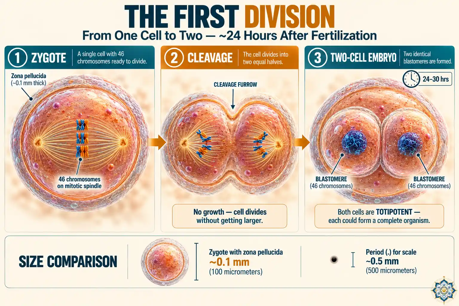
No growth phase. In a typical cell division, the cell grows larger before splitting. The zygote doesn’t. It divides without increasing in volume, producing two cells (called blastomeres) that are each half the size of the original. This pattern continues — the embryo gets more cells but not bigger — until it implants in the uterus days later. About two weeks after that, the single most consequential event of your embryonic life occurs: gastrulation, when the flat embryonic disc transforms into the three germ layers that will build every organ in your body.
Genome mixing. This first division is when the two parental genomes are truly combined for the first time. The 46 chromosomes (23 from each parent) are replicated together, lined up on the same spindle, and pulled apart into two complete sets. Each daughter cell receives a full copy of the new combined genome.
Totipotency. Both of these first two cells are totipotent — each one alone could theoretically develop into a complete human being. This is how identical twins can form: if these two cells separate, each can produce a full embryo. By the 8–16 cell stage, this totipotency begins to fade. This gradual loss of flexibility continues throughout development — in the first two trimesters, the fetus can even heal skin wounds without any scar at all, an ability it loses before birth and never fully recovers (how wounds heal and why scars form).
Error-prone. This first division has a surprisingly high error rate. Studies of IVF embryos show that 50–80% of human embryos have chromosomal abnormalities by the time they reach the 8-cell stage. Most of these errors are fatal, which is a major reason why more than 50% of all fertilized eggs never result in a pregnancy. Human reproduction is, by engineering standards, remarkably inefficient.
10. What Happens After Fertilization — Minute-by-Minute Timeline
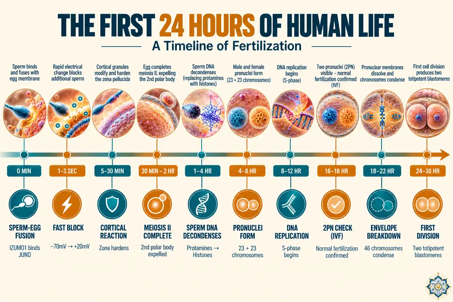
Here’s the complete picture of what happens after fertilization, condensed into a single reference table:
| Time after sperm entry | Event | What’s happening |
|---|---|---|
| 0 seconds | Sperm-egg membrane fusion | IZUMO1-JUNO binding; sperm contents enter egg |
| 1–3 seconds | Fast block to polyspermy | Membrane depolarization from –70mV to +20mV |
| 30 seconds – 5 min | Calcium wave | Oscillating calcium signals sweep across egg; trigger cortical reaction |
| 5–30 minutes | Cortical reaction | Cortical granules release enzymes; zona pellucida hardens |
| 30–40 minutes | JUNO shedding | JUNO receptor removed from egg surface |
| 30 min – 2 hours | Meiosis II completion | Egg finishes its final division; second polar body expelled |
| 1–4 hours | Sperm DNA decondensation | Protamines replaced by histones; sperm DNA unwinds |
| 4–8 hours | Pronuclei formation | Male and female pronuclei become visible |
| 6–12 hours | DNA demethylation begins | Epigenetic reprogramming of paternal DNA |
| 8–12 hours | DNA replication (S-phase) | Both pronuclei replicate their DNA |
| 16–18 hours | 2PN stage (IVF checkpoint) | Two pronuclei visible side-by-side; confirms normal fertilization |
| 18–22 hours | Pronuclear envelope breakdown | Nuclear membranes dissolve; chromosomes condense |
| 20–24 hours | First mitotic spindle forms | 46 chromosomes align together for the first time |
| 24–30 hours | First cleavage division | One cell becomes two blastomeres; each has 46 chromosomes |
11. What IVF Teaches Us About Fertilization
Much of what we know about what happens after fertilization comes from in vitro fertilization (IVF), where the process occurs in a dish under controlled conditions. IVF has given embryologists a front-row seat to processes that normally occur inside the fallopian tube, invisible to observation.
Several key insights have come from IVF:
Timing matters more than quantity. In conventional IVF, eggs are incubated with thousands of sperm. But since the development of ICSI (intracytoplasmic sperm injection) in 1992, embryologists learned that a single sperm injected directly into the egg can produce a healthy embryo — proving that the hundreds of sperm surrounding the egg in natural fertilization aren’t all necessary for the process itself. They’re needed to get there, not to do the job.
The 2PN check. At 16–18 hours after insemination, IVF embryologists check for two pronuclei. Zero pronuclei means fertilization failed. One pronucleus suggests the egg activated without being fertilized (parthenogenesis). Three pronuclei means two sperm entered (polyspermy). Only embryos showing exactly two pronuclei are considered normally fertilized.
Time-lapse imaging. Modern IVF labs use time-lapse incubators that photograph embryos every 5–10 minutes without disturbing them. This technology has revealed that the exact timing of division events predicts embryo quality — embryos that divide too fast or too slow are less likely to develop normally. The first division should happen between 25–27 hours after ICSI, and deviations of even a few hours correlate with lower implantation rates.
Failure is normal. IVF data has shown that even under ideal laboratory conditions, a significant percentage of embryos stop developing by day 3–5. This matches what happens naturally — most fertilized eggs never become pregnancies. The leading cause is aneuploidy (an abnormal chromosome count), which can result from errors during the egg’s meiosis (often related to maternal age) or during the early cleavage divisions.
A number worth knowing: In women under 35, about 40–50% of embryos created through IVF are chromosomally normal. In women over 40, that figure drops to 10–20%. This is because eggs, which are formed before a woman is born, accumulate errors in their chromosome-separation machinery over decades. It’s one of the clearest examples of biological aging at the cellular level. Learn more at ASRM’s IVF resource page.
Frequently asked questions
How long does human fertilization actually take?
The entire process — from the sperm binding the egg’s outer coat (zona pellucida) to the completion of the first mitotic division — takes about 24 hours. Sperm penetration itself takes roughly 20 minutes. Pronuclei form over 6–8 hours and merge over the next few hours. The first cell division typically happens around 24–30 hours after the sperm enters the egg.
How many sperm actually reach the egg?
Out of roughly 200–300 million sperm released during ejaculation, only about 200 reach the egg in the fallopian tube. That’s a survival rate of less than 0.0001%. Most are filtered out by cervical mucus, destroyed by the acidic vaginal environment, or swim into the wrong fallopian tube.
What is the cortical reaction?
The cortical reaction is the egg’s mechanism for blocking additional sperm after one has entered. Within seconds of sperm entry, cortical granules just below the egg’s surface release enzymes that harden the zona pellucida — the egg’s outer coat. This prevents polyspermy, which would give the embryo too many chromosomes and be fatal.
What is a pronucleus?
A pronucleus is a haploid nucleus — one containing 23 chromosomes — found in the fertilized egg before the parental genomes merge. The egg’s nucleus becomes the female pronucleus, and the sperm’s DNA decondenses to form the male pronucleus. These two pronuclei migrate toward each other and their chromosomes combine during the first cell division.
What is the acrosome reaction?
The acrosome reaction is the release of digestive enzymes from the cap-like structure (acrosome) at the tip of the sperm head. When the sperm binds to ZP3 glycoprotein on the zona pellucida, the acrosome releases hyaluronidase and acrosin, which digest a path through the egg’s outer layers so the sperm can reach the egg membrane.
When does DNA from mother and father actually combine?
The parental genomes don’t fuse the way most people imagine. The two pronuclei migrate toward the center of the egg, their nuclear envelopes break down, and the 46 chromosomes (23 from each parent) line up together on the first mitotic spindle. The two sets of DNA are first truly combined when they are replicated and separated during this first cell division, around 24 hours after sperm entry.
What decides whether the baby will be male or female?
Biological sex is determined by the sperm. Every egg carries an X chromosome. Sperm carry either an X or a Y chromosome. If an X-bearing sperm fertilizes the egg, the result is XX (female). If a Y-bearing sperm fertilizes it, the result is XY (male). This is decided at the instant of fertilization and cannot change afterward.
Can more than one sperm enter the egg?
Normally, no. The egg has two blocking mechanisms — a fast electrical block that changes the egg’s membrane potential within seconds, and a slower cortical reaction that permanently hardens the outer coat. If these mechanisms fail and two sperm enter (polyspermy), the resulting cell has 69 chromosomes instead of 46, which is almost always fatal. Polyspermy occurs in less than 1% of human fertilizations.
What is capacitation?
Capacitation is a series of biochemical changes that sperm undergo inside the female reproductive tract over 7–10 hours. These changes destabilize the sperm’s membrane, enable the acrosome reaction, and give the sperm a hyperactivated whip-like swimming motion. Sperm that have not been capacitated cannot fertilize an egg.
What happens to the other sperm that don’t fertilize the egg?
The vast majority of sperm die within hours to days inside the female reproductive tract. Some are destroyed by the acidic vaginal pH, others are filtered by cervical mucus, and many are engulfed by white blood cells. The few hundred that reach the egg but fail to penetrate it eventually degrade and are reabsorbed by the body. Sperm can survive in the reproductive tract for up to 5 days under favorable conditions.
References
- Alberts, B., Johnson, A., Lewis, J., et al. Molecular Biology of the Cell, 6th Edition. Garland Science, 2014. Chapter 21: Sexual Reproduction.
- Gilbert, S.F. Developmental Biology, 12th Edition. Sinauer Associates, 2019. Chapters 7–8: Fertilization.
- Bianchi, E., Doe, B., Goulding, D., Wright, G.J. “Juno is the egg Izumo receptor and is essential for mammalian fertilization.” Nature, 508, 483–487 (2014).
- MITOMAP: A Human Mitochondrial Genome Database. mitomap.org/MITOMAP.
- Lundin, K., Bergh, C., Hardarson, T. “Early embryo cleavage is a strong indicator of embryo quality in human IVF.” Human Reproduction, 16(12), 2652–2657 (2001).
- Franasiak, J.M., et al. “The nature of aneuploidy with increasing age of the female partner.” Fertility and Sterility, 101(3), 656–663 (2014).
- American Society for Reproductive Medicine. In Vitro Fertilization (IVF): What is it? asrm.org.
- Mayer, W., Niveleau, A., Walter, J., Fundele, R., Haaf, T. “Demethylation of the zygotic paternal genome.” Nature, 403, 501–502 (2000).
- Clift, D., Schuh, M. “Restarting life: fertilization and the transition from meiosis to mitosis.” Nature Reviews Molecular Cell Biology, 14, 549–562 (2013).
This article is for educational purposes. It provides a science-based overview of human fertilization and early embryo development. HowLifeBuilds is an independent science publication. Content is reviewed for accuracy but does not substitute for professional medical or reproductive health advice. If you have questions about fertility or reproductive health, consult a qualified healthcare provider.
Follow HowLifeBuilds
@howlifebuilds